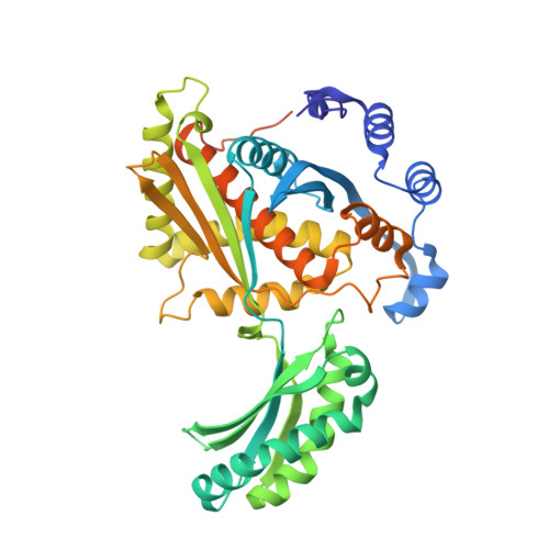
5WPK

Structure of the class II 3-hydroxy-3-methylglutaryl-CoA reductase from Streptococcus pneumoniae bound to HMG-CoA and in a partially closed conformation


Experimental Data Snapshot

  • Method: X-RAY DIFFRACTION
  • Resolution: 2.30 Å
  • R-Value Free: 0.233 
  • R-Value Work: 0.177 
  • R-Value Observed: 0.187 

Starting Model: experimental
View more details

wwPDB Validation   3D Report Full Report


Ligand Structure Quality Assessment 


This is version 1.2 of the entry. See complete history


Literature

Structural Features and Domain Movements Controlling Substrate Binding and Cofactor Specificity in Class II HMG-CoA Reductase.

Miller, B.R.Kung, Y.

(2018) Biochemistry 57: 654-662

  • DOI: https://doi.org/10.1021/acs.biochem.7b00999
  • Primary Citation of Related Structures:  
    5WPJ, 5WPK

  • PubMed Abstract: 

    The key mevalonate pathway enzyme 3-hydroxy-3-methylglutaryl coenzyme A (HMG-CoA) reductase (HMGR) uses the cofactor NAD(P)H to reduce HMG-CoA to mevalonate in the production of countless metabolites and natural products. Although inhibition of HMGR by statin drugs is well-understood, several mechanistic details of HMGR catalysis remain unresolved, and the structural basis for the wide range of cofactor specificity for either NADH or NADPH among HMGRs from different organisms is also unknown. Here, we present crystal structures of HMGR from Streptococcus pneumoniae (SpHMGR) alongside kinetic data of the enzyme's cofactor preferences. Our structure of SpHMGR bound with its kinetically preferred NADPH cofactor suggests how NADPH-specific binding and recognition are achieved. In addition, our structure of HMG-CoA-bound SpHMGR reveals large, previously unknown conformational domain movements that may control HMGR substrate binding and enable cofactor exchange without intermediate release during the catalytic cycle. Taken together, this work provides critical new insights into both the HMGR reaction mechanism and the structural basis of cofactor specificity.


  • Organizational Affiliation

    Department of Chemistry, Bryn Mawr College , 101 North Merion Avenue, Bryn Mawr, Pennsylvania 19010, United States.


Macromolecules
Find similar proteins by:  (by identity cutoff)  |  3D Structure
Entity ID: 1
MoleculeChains Sequence LengthOrganismDetailsImage
3-hydroxy-3-methylglutaryl coenzyme A reductase
A, B
426Streptococcus pneumoniaeMutation(s): 0 
Gene Names: 
EC: 1.1.1.34 (PDB Primary Data), 1.1.1.88 (UniProt)
UniProt
Find proteins for Q8DNS5 (Streptococcus pneumoniae (strain ATCC BAA-255 / R6))
Explore Q8DNS5 
Go to UniProtKB:  Q8DNS5
Entity Groups  
Sequence Clusters30% Identity50% Identity70% Identity90% Identity95% Identity100% Identity
UniProt GroupQ8DNS5
Sequence Annotations
Expand
  • Reference Sequence
Small Molecules
Ligands 3 Unique
IDChains Name / Formula / InChI Key2D Diagram3D Interactions
HMG
Query on HMG

Download Ideal Coordinates CCD File 
C [auth A],
F [auth B]
3-HYDROXY-3-METHYLGLUTARYL-COENZYME A
C27 H39 N7 O20 P3 S
CABVTRNMFUVUDM-VRHQGPGLSA-I
PE4
Query on PE4

Download Ideal Coordinates CCD File 
E [auth B],
I [auth B]
2-{2-[2-(2-{2-[2-(2-ETHOXY-ETHOXY)-ETHOXY]-ETHOXY}-ETHOXY)-ETHOXY]-ETHOXY}-ETHANOL
C16 H34 O8
PJWQOENWHPEPKI-UHFFFAOYSA-N
GOL
Query on GOL

Download Ideal Coordinates CCD File 
D [auth A],
G [auth B],
H [auth B]
GLYCEROL
C3 H8 O3
PEDCQBHIVMGVHV-UHFFFAOYSA-N
Experimental Data & Validation

Experimental Data

  • Method: X-RAY DIFFRACTION
  • Resolution: 2.30 Å
  • R-Value Free: 0.233 
  • R-Value Work: 0.177 
  • R-Value Observed: 0.187 
  • Space Group: P 1 21 1
Unit Cell:
Length ( Å )Angle ( ˚ )
a = 57.865α = 90
b = 131.248β = 102.54
c = 57.957γ = 90
Software Package:
Software NamePurpose
PHENIXrefinement
iMOSFLMdata reduction
Aimlessdata scaling
PHENIXphasing

Structure Validation

View Full Validation Report


Ligand Structure Quality Assessment 


Entry History & Funding Information

Deposition Data


Funding OrganizationLocationGrant Number
National Institutes of Health/National Institute of General Medical Sciences (NIH/NIGMS)United StatesGM116029

Revision History  (Full details and data files)

  • Version 1.0: 2018-04-25
    Type: Initial release
  • Version 1.1: 2020-01-01
    Changes: Author supporting evidence
  • Version 1.2: 2023-10-04
    Changes: Data collection, Database references, Refinement description