6GSZ | pdb_00006gsz

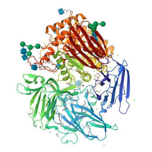
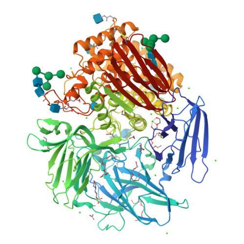
Crystal structure of native alfa-L-rhamnosidase from Aspergillus terreus


Experimental Data Snapshot

  • Method: X-RAY DIFFRACTION
  • Resolution: 1.38 Å
  • R-Value Free: 
    0.168 (Depositor), 0.169 (DCC) 
  • R-Value Work: 
    0.134 (Depositor), 0.138 (DCC) 
  • R-Value Observed: 
    0.136 (Depositor) 

wwPDB Validation   3D Report Full Report


Ligand Structure Quality Assessment 

Created with Raphaël 2.3.0Worse 01 BetterLigand structure goodness of fit to experimental dataBest fitted NAGClick on this verticalbar to view detailsBest fitted MPOClick on this verticalbar to view detailsBest fitted BGCClick on this verticalbar to view detailsBest fitted DTUClick on this verticalbar to view details

This is version 2.1 of the entry. See complete history


Literature

Crystal structure of native alpha-L-rhamnosidase from Aspergillus terreus.

Pachl, P.Skerlova, J.Simcikova, D.Kotik, M.Krenkova, A.Mader, P.Brynda, J.Kapesova, J.Kren, V.Otwinowski, Z.Rezacova, P.

(2018) Acta Crystallogr D Struct Biol 74: 1078-1084

  • DOI: https://doi.org/10.1107/S2059798318013049
  • Primary Citation of Related Structures:  
    6GSZ

  • PubMed Abstract: 

    α-L-Rhamnosidases cleave terminal nonreducing α-L-rhamnosyl residues from many natural rhamnoglycosides. This makes them catalysts of interest for various biotechnological applications. The X-ray structure of the GH78 family α-L-rhamnosidase from Aspergillus terreus has been determined at 1.38 Å resolution using the sulfur single-wavelength anomalous dispersion phasing method. The protein was isolated from its natural source in the native glycosylated form, and the active site contained a glucose molecule, probably from the growth medium. In addition to its catalytic domain, the α-L-rhamnosidase from A. terreus contains four accessory domains of unknown function. The structural data suggest that two of these accessory domains, E and F, might play a role in stabilizing the aglycon portion of the bound substrate.


  • Organizational Affiliation

    Structural Biology, Institute of Organic Chemistry and Biochemistry, The Czech Academy of Sciences, Flemingovo nám. 2, 16610 Prague 6, Czech Republic.


Macromolecules
Find similar proteins by:  (by identity cutoff)  |  3D Structure
Entity ID: 1
MoleculeChains Sequence LengthOrganismDetailsImage
Alpha-L-rhamnosidase870Aspergillus terreusMutation(s): 0 
Gene Names: alpha-Rha
EC: 3.2.1.40
UniProt
Find proteins for I0AZ41 (Aspergillus terreus)
Explore I0AZ41 
Go to UniProtKB:  I0AZ41
Entity Groups  
Sequence Clusters30% Identity50% Identity70% Identity90% Identity95% Identity100% Identity
UniProt GroupI0AZ41
Glycosylation
Glycosylation Sites: 7
Sequence Annotations
Expand
  • Reference Sequence
Oligosaccharides

Help

Entity ID: 2
MoleculeChains Length2D Diagram Glycosylation3D Interactions
alpha-D-mannopyranose-(1-3)-beta-D-mannopyranose-(1-4)-2-acetamido-2-deoxy-beta-D-glucopyranose-(1-4)-2-acetamido-2-deoxy-beta-D-glucopyranose
B
4N-Glycosylation
Glycosylation Resources
GlyTouCan:  G81315DD
GlyCosmos:  G81315DD
GlyGen:  G81315DD
Entity ID: 3
MoleculeChains Length2D Diagram Glycosylation3D Interactions
alpha-D-mannopyranose-(1-3)-[alpha-D-mannopyranose-(1-6)]alpha-D-mannopyranose-(1-6)-[alpha-D-mannopyranose-(1-3)]beta-D-mannopyranose-(1-4)-2-acetamido-2-deoxy-beta-D-glucopyranose-(1-4)-2-acetamido-2-deoxy-beta-D-glucopyranose
C
7N-Glycosylation
Glycosylation Resources
GlyTouCan:  G55220VL
GlyCosmos:  G55220VL
GlyGen:  G55220VL
Entity ID: 4
MoleculeChains Length2D Diagram Glycosylation3D Interactions
alpha-D-mannopyranose-(1-3)-[alpha-D-mannopyranose-(1-6)]beta-D-mannopyranose-(1-4)-2-acetamido-2-deoxy-beta-D-glucopyranose-(1-4)-2-acetamido-2-deoxy-beta-D-glucopyranose
D
5N-Glycosylation
Glycosylation Resources
GlyTouCan:  G22768VO
GlyCosmos:  G22768VO
GlyGen:  G22768VO
Entity ID: 5
MoleculeChains Length2D Diagram Glycosylation3D Interactions
2-acetamido-2-deoxy-beta-D-glucopyranose-(1-4)-2-acetamido-2-deoxy-beta-D-glucopyranose
E
2N-Glycosylation
Glycosylation Resources
GlyTouCan:  G42666HT
GlyCosmos:  G42666HT
GlyGen:  G42666HT
Entity ID: 6
MoleculeChains Length2D Diagram Glycosylation3D Interactions
alpha-D-mannopyranose-(1-3)-[alpha-D-mannopyranose-(1-6)]alpha-D-mannopyranose-(1-6)-beta-D-mannopyranose-(1-4)-2-acetamido-2-deoxy-beta-D-glucopyranose-(1-4)-2-acetamido-2-deoxy-beta-D-glucopyranose
F
6N-Glycosylation
Glycosylation Resources
GlyTouCan:  G94106MV
GlyCosmos:  G94106MV
GlyGen:  G94106MV
Small Molecules
Ligands 11 Unique
IDChains Name / Formula / InChI Key2D Diagram3D Interactions
1PE
Query on 1PE

Download Ideal Coordinates CCD File 
DA [auth A],
JA [auth A],
KA [auth A]
PENTAETHYLENE GLYCOL
C10 H22 O6
JLFNLZLINWHATN-UHFFFAOYSA-N
NAG
Query on NAG

Download Ideal Coordinates CCD File 
G [auth A],
H [auth A]
2-acetamido-2-deoxy-beta-D-glucopyranose
C8 H15 N O6
OVRNDRQMDRJTHS-FMDGEEDCSA-N
MPO
Query on MPO

Download Ideal Coordinates CCD File 
WA [auth A]3[N-MORPHOLINO]PROPANE SULFONIC ACID
C7 H15 N O4 S
DVLFYONBTKHTER-UHFFFAOYSA-N
PG4
Query on PG4

Download Ideal Coordinates CCD File 
EA [auth A]TETRAETHYLENE GLYCOL
C8 H18 O5
UWHCKJMYHZGTIT-UHFFFAOYSA-N
BGC
Query on BGC

Download Ideal Coordinates CCD File 
XA [auth A]beta-D-glucopyranose
C6 H12 O6
WQZGKKKJIJFFOK-VFUOTHLCSA-N
DTU
Query on DTU

Download Ideal Coordinates CCD File 
PA [auth A](2R,3S)-1,4-DIMERCAPTOBUTANE-2,3-DIOL
C4 H10 O2 S2
VHJLVAABSRFDPM-ZXZARUISSA-N
PGE
Query on PGE

Download Ideal Coordinates CCD File 
FA [auth A],
IA [auth A],
LA [auth A],
OA [auth A]
TRIETHYLENE GLYCOL
C6 H14 O4
ZIBGPFATKBEMQZ-UHFFFAOYSA-N
PG0
Query on PG0

Download Ideal Coordinates CCD File 
GA [auth A],
HA [auth A],
MA [auth A]
2-(2-METHOXYETHOXY)ETHANOL
C5 H12 O3
SBASXUCJHJRPEV-UHFFFAOYSA-N
EDO
Query on EDO

Download Ideal Coordinates CCD File 
NA [auth A]1,2-ETHANEDIOL
C2 H6 O2
LYCAIKOWRPUZTN-UHFFFAOYSA-N
ACT
Query on ACT

Download Ideal Coordinates CCD File 
QA [auth A]
RA [auth A]
SA [auth A]
TA [auth A]
UA [auth A]
ACETATE ION
C2 H3 O2
QTBSBXVTEAMEQO-UHFFFAOYSA-M
CA
Query on CA

Download Ideal Coordinates CCD File 
AA [auth A]
BA [auth A]
CA [auth A]
I [auth A]
J [auth A]
CALCIUM ION
Ca
BHPQYMZQTOCNFJ-UHFFFAOYSA-N
Experimental Data & Validation

Experimental Data

  • Method: X-RAY DIFFRACTION
  • Resolution: 1.38 Å
  • R-Value Free:  0.168 (Depositor), 0.169 (DCC) 
  • R-Value Work:  0.134 (Depositor), 0.138 (DCC) 
  • R-Value Observed: 0.136 (Depositor) 
Space Group: P 21 21 21
Unit Cell:
Length ( Å )Angle ( ˚ )
a = 90.26α = 90
b = 106.248β = 90
c = 119.832γ = 90
Software Package:
Software NamePurpose
REFMACrefinement
HKL-2000data reduction
HKL-2000data scaling
MLPHAREphasing

Structure Validation

View Full Validation Report


Ligand Structure Quality Assessment 

Created with Raphaël 2.3.0Worse 01 BetterLigand structure goodness of fit to experimental dataBest fitted NAGClick on this verticalbar to view detailsBest fitted MPOClick on this verticalbar to view detailsBest fitted BGCClick on this verticalbar to view detailsBest fitted DTUClick on this verticalbar to view details

Entry History & Funding Information

Deposition Data


Funding OrganizationLocationGrant Number
Ministry of Education (Czech Republic)Czech RepublicLO1304

Revision History  (Full details and data files)

  • Version 1.0: 2018-11-14
    Type: Initial release
  • Version 2.0: 2020-07-29
    Type: Remediation
    Reason: Carbohydrate remediation
    Changes: Advisory, Atomic model, Data collection, Derived calculations, Structure summary
  • Version 2.1: 2024-11-20
    Changes: Data collection, Database references, Derived calculations, Structure summary