6HAF | pdb_00006haf

Pyruvate oxidase variant E59Q from L. plantarum in complex with phosphate


Experimental Data Snapshot

  • Method: X-RAY DIFFRACTION
  • Resolution: 1.30 Å
  • R-Value Free: 
    0.156 (Depositor), 0.160 (DCC) 
  • R-Value Work: 
    0.134 (Depositor), 0.140 (DCC) 
  • R-Value Observed: 
    0.135 (Depositor) 

wwPDB Validation   3D Report Full Report


Ligand Structure Quality Assessment 

Created with Raphaël 2.3.0Worse 01 BetterLigand structure goodness of fit to experimental dataBest fitted TPPClick on this verticalbar to view details

This is version 2.1 of the entry. See complete history


Literature

Low-barrier hydrogen bonds in enzyme cooperativity.

Dai, S.Funk, L.M.von Pappenheim, F.R.Sautner, V.Paulikat, M.Schroder, B.Uranga, J.Mata, R.A.Tittmann, K.

(2019) Nature 573: 609-613

  • DOI: https://doi.org/10.1038/s41586-019-1581-9
  • Primary Citation of Related Structures:  
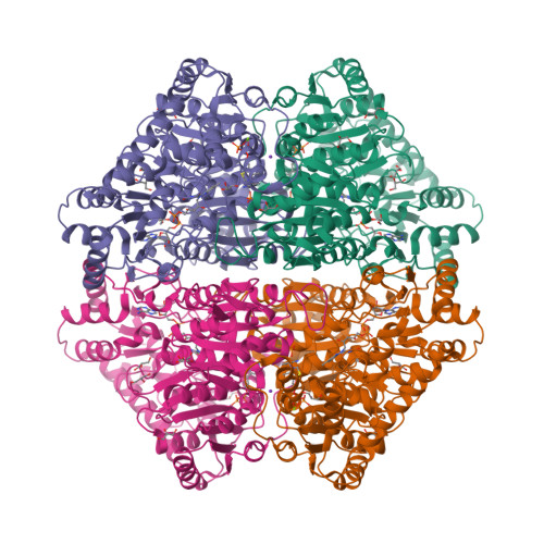
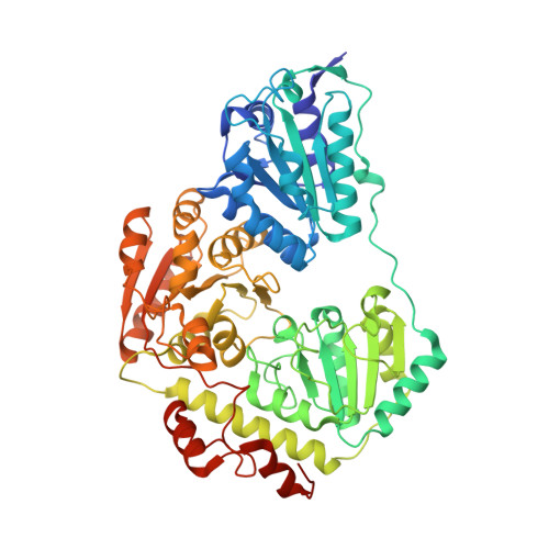
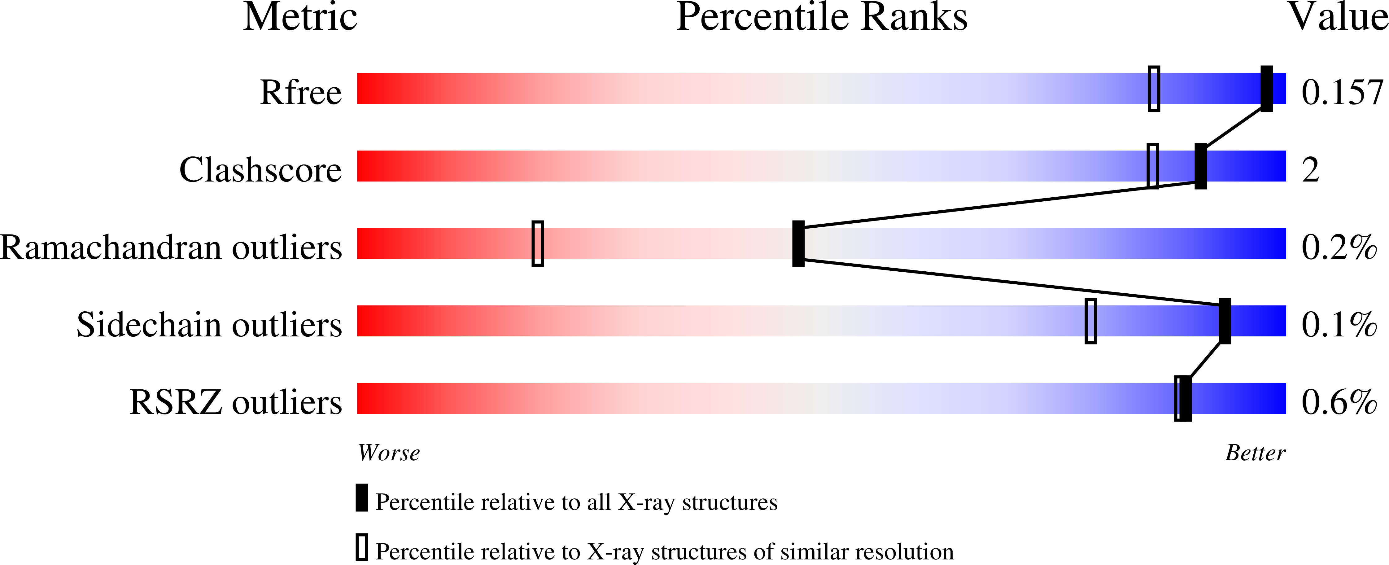
    6HA3, 6HAD, 6HAF, 6RJB, 6RJC

  • PubMed Abstract: 

    The underlying molecular mechanisms of cooperativity and allosteric regulation are well understood for many proteins, with haemoglobin and aspartate transcarbamoylase serving as prototypical examples 1,2 . The binding of effectors typically causes a structural transition of the protein that is propagated through signalling pathways to remote sites and involves marked changes on the tertiary and sometimes even the quaternary level 1-5 . However, the origin of these signals and the molecular mechanism of long-range signalling at an atomic level remain unclear 5-8 . The different spatial scales and timescales in signalling pathways render experimental observation challenging; in particular, the positions and movement of mobile protons cannot be visualized by current methods of structural analysis. Here we report the experimental observation of fluctuating low-barrier hydrogen bonds as switching elements in cooperativity pathways of multimeric enzymes. We have observed these low-barrier hydrogen bonds in ultra-high-resolution X-ray crystallographic structures of two multimeric enzymes, and have validated their assignment using computational calculations. Catalytic events at the active sites switch between low-barrier hydrogen bonds and ordinary hydrogen bonds in a circuit that consists of acidic side chains and water molecules, transmitting a signal through the collective repositioning of protons by behaving as an atomistic Newton's cradle. The resulting communication synchronizes catalysis in the oligomer. Our studies provide several lines of evidence and a working model for not only the existence of low-barrier hydrogen bonds in proteins, but also a connection to enzyme cooperativity. This finding suggests new principles of drug and enzyme design, in which sequences of residues can be purposefully included to enable long-range communication and thus the regulation of engineered biomolecules.


  • Organizational Affiliation

    Department of Molecular Enzymology, Göttingen Centre for Molecular Biosciences and Albrecht-von-Haller Institute, Georg-August University Göttingen, Göttingen, Germany.


Macromolecules
Find similar proteins by:  (by identity cutoff)  |  3D Structure
Entity ID: 1
MoleculeChains Sequence LengthOrganismDetailsImage
Pyruvate oxidase
A, B
603Lactiplantibacillus plantarumMutation(s): 1 
Gene Names: spxBC4O30_12890CUR48_03890
EC: 1.2.3.3
UniProt
Find proteins for P37063 (Lactiplantibacillus plantarum (strain ATCC BAA-793 / NCIMB 8826 / WCFS1))
Explore P37063 
Go to UniProtKB:  P37063
Entity Groups  
Sequence Clusters30% Identity50% Identity70% Identity90% Identity95% Identity100% Identity
UniProt GroupP37063
Sequence Annotations
Expand
  • Reference Sequence
Small Molecules
Ligands 6 Unique
IDChains Name / Formula / InChI Key2D Diagram3D Interactions
FAD
Query on FAD

Download Ideal Coordinates CCD File 
C [auth A],
O [auth B]
FLAVIN-ADENINE DINUCLEOTIDE
C27 H33 N9 O15 P2
VWWQXMAJTJZDQX-UYBVJOGSSA-N
TPP (Subject of Investigation/LOI)
Query on TPP

Download Ideal Coordinates CCD File 
F [auth A],
R [auth B]
THIAMINE DIPHOSPHATE
C12 H19 N4 O7 P2 S
AYEKOFBPNLCAJY-UHFFFAOYSA-O
PO4
Query on PO4

Download Ideal Coordinates CCD File 
N [auth A],
X [auth B]
PHOSPHATE ION
O4 P
NBIIXXVUZAFLBC-UHFFFAOYSA-K
GOL
Query on GOL

Download Ideal Coordinates CCD File 
G [auth A]
H [auth A]
I [auth A]
J [auth A]
K [auth A]
GLYCEROL
C3 H8 O3
PEDCQBHIVMGVHV-UHFFFAOYSA-N
K
Query on K

Download Ideal Coordinates CCD File 
D [auth A],
P [auth B]
POTASSIUM ION
K
NPYPAHLBTDXSSS-UHFFFAOYSA-N
MG
Query on MG

Download Ideal Coordinates CCD File 
E [auth A],
Q [auth B]
MAGNESIUM ION
Mg
JLVVSXFLKOJNIY-UHFFFAOYSA-N
Experimental Data & Validation

Experimental Data

  • Method: X-RAY DIFFRACTION
  • Resolution: 1.30 Å
  • R-Value Free:  0.156 (Depositor), 0.160 (DCC) 
  • R-Value Work:  0.134 (Depositor), 0.140 (DCC) 
  • R-Value Observed: 0.135 (Depositor) 
Space Group: C 2 2 21
Unit Cell:
Length ( Å )Angle ( ˚ )
a = 119.18α = 90
b = 154.35β = 90
c = 165.52γ = 90
Software Package:
Software NamePurpose
XSCALEdata scaling
PHENIXrefinement
PDB_EXTRACTdata extraction
XDSdata reduction
PHENIXphasing

Structure Validation

View Full Validation Report


Ligand Structure Quality Assessment 

Created with Raphaël 2.3.0Worse 01 BetterLigand structure goodness of fit to experimental dataBest fitted TPPClick on this verticalbar to view details

Entry History & Funding Information

Deposition Data


Funding OrganizationLocationGrant Number
German Research FoundationGermanyFOR 1296/TP3

Revision History  (Full details and data files)

  • Version 1.0: 2019-08-21
    Type: Initial release
  • Version 1.1: 2019-09-18
    Changes: Data collection, Database references
  • Version 1.2: 2019-10-02
    Changes: Data collection, Database references
  • Version 1.3: 2019-10-09
    Changes: Data collection, Database references
  • Version 2.0: 2021-08-04
    Changes: Atomic model, Author supporting evidence, Data collection, Derived calculations, Non-polymer description, Structure summary
  • Version 2.1: 2024-05-15
    Changes: Data collection, Database references