6WHV

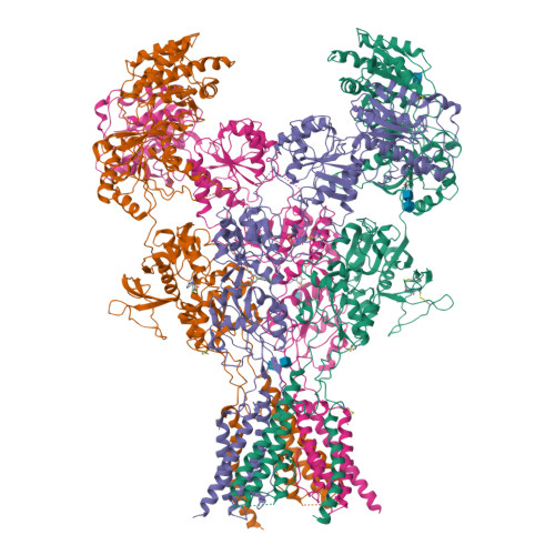
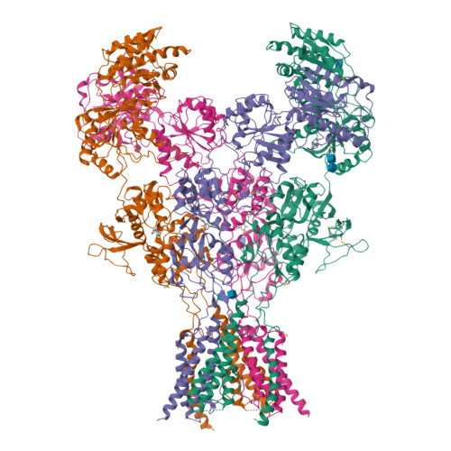
GluN1b-GluN2B NMDA receptor in complex with SDZ 220-040 and L689,560, class 2


Experimental Data Snapshot

  • Method: ELECTRON MICROSCOPY
  • Resolution: 4.05 Å
  • Aggregation State: PARTICLE 
  • Reconstruction Method: SINGLE PARTICLE 

wwPDB Validation   3D Report Full Report


This is version 1.1 of the entry. See complete history


Literature

Structural Basis of Functional Transitions in Mammalian NMDA Receptors.

Chou, T.H.Tajima, N.Romero-Hernandez, A.Furukawa, H.

(2020) Cell 182: 357-371.e13

  • DOI: https://doi.org/10.1016/j.cell.2020.05.052
  • Primary Citation of Related Structures:  
    6USU, 6USV, 6WHR, 6WHS, 6WHT, 6WHU, 6WHV, 6WHW, 6WHX, 6WHY, 6WI0, 6WI1

  • PubMed Abstract: 

    Excitatory neurotransmission meditated by glutamate receptors including N-methyl-D-aspartate receptors (NMDARs) is pivotal to brain development and function. NMDARs are heterotetramers composed of GluN1 and GluN2 subunits, which bind glycine and glutamate, respectively, to activate their ion channels. Despite importance in brain physiology, the precise mechanisms by which activation and inhibition occur via subunit-specific binding of agonists and antagonists remain largely unknown. Here, we show the detailed patterns of conformational changes and inter-subunit and -domain reorientation leading to agonist-gating and subunit-dependent competitive inhibition by providing multiple structures in distinct ligand states at 4 Å or better. The structures reveal that activation and competitive inhibition by both GluN1 and GluN2 antagonists occur by controlling the tension of the linker between the ligand-binding domain and the transmembrane ion channel of the GluN2 subunit. Our results provide detailed mechanistic insights into NMDAR pharmacology, activation, and inhibition, which are fundamental to the brain physiology.


  • Organizational Affiliation

    WM Keck Structural Biology Laboratory, Cold Spring Harbor Laboratory, Cold Spring Harbor, NY 11724, USA.


Macromolecules
Find similar proteins by:  (by identity cutoff)  |  3D Structure
Entity ID: 1
MoleculeChains Sequence LengthOrganismDetailsImage
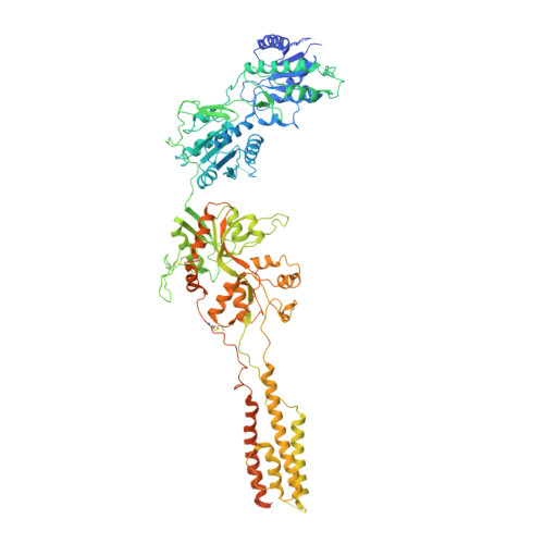
Glutamate receptor ionotropic, NMDA 1
A, C
959Rattus norvegicusMutation(s): 0 
Gene Names: Grin1Nmdar1
Membrane Entity: Yes 
UniProt
Find proteins for P35439 (Rattus norvegicus)
Explore P35439 
Go to UniProtKB:  P35439
Entity Groups  
Sequence Clusters30% Identity50% Identity70% Identity90% Identity95% Identity100% Identity
UniProt GroupP35439
Glycosylation
Glycosylation Sites: 1Go to GlyGen: P35439-1
Sequence Annotations
Expand
  • Reference Sequence
Find similar proteins by:  (by identity cutoff)  |  3D Structure
Entity ID: 2
MoleculeChains Sequence LengthOrganismDetailsImage
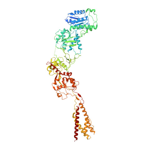
Glutamate receptor ionotropic, NMDA 2B
B, D
883Rattus norvegicusMutation(s): 0 
Gene Names: Grin2b
Membrane Entity: Yes 
UniProt
Find proteins for Q00960 (Rattus norvegicus)
Explore Q00960 
Go to UniProtKB:  Q00960
Entity Groups  
Sequence Clusters30% Identity50% Identity70% Identity90% Identity95% Identity100% Identity
UniProt GroupQ00960
Glycosylation
Glycosylation Sites: 2Go to GlyGen: Q00960-1
Sequence Annotations
Expand
  • Reference Sequence
Oligosaccharides

Help

Entity ID: 3
MoleculeChains Length2D Diagram Glycosylation3D Interactions
2-acetamido-2-deoxy-beta-D-glucopyranose-(1-4)-2-acetamido-2-deoxy-beta-D-glucopyranose
E
2N-Glycosylation
Glycosylation Resources
GlyTouCan:  G42666HT
GlyCosmos:  G42666HT
GlyGen:  G42666HT
Small Molecules
Ligands 3 Unique
IDChains Name / Formula / InChI Key2D Diagram3D Interactions
QGP (Subject of Investigation/LOI)
Query on QGP

Download Ideal Coordinates CCD File 
G [auth B],
J [auth D]
(2S)-2-amino-3-[2',4'-dichloro-4-hydroxy-5-(phosphonomethyl)biphenyl-3-yl]propanoic acid
C16 H16 Cl2 N O6 P
NYZFUZCCDOSQBG-AWEZNQCLSA-N
QGM (Subject of Investigation/LOI)
Query on QGM

Download Ideal Coordinates CCD File 
I [auth C](2R,4S)-5,7-dichloro-4-[(phenylcarbamoyl)amino]-1,2,3,4-tetrahydroquinoline-2-carboxylic acid
C17 H15 Cl2 N3 O3
UCKHICKHGAOGAP-UONOGXRCSA-N
NAG
Query on NAG

Download Ideal Coordinates CCD File 
F [auth A],
H [auth B],
K [auth D],
L [auth D]
2-acetamido-2-deoxy-beta-D-glucopyranose
C8 H15 N O6
OVRNDRQMDRJTHS-FMDGEEDCSA-N
Experimental Data & Validation

Experimental Data

  • Method: ELECTRON MICROSCOPY
  • Resolution: 4.05 Å
  • Aggregation State: PARTICLE 
  • Reconstruction Method: SINGLE PARTICLE 
EM Software:
TaskSoftware PackageVersion
RECONSTRUCTIONcisTEM1.0.0

Structure Validation

View Full Validation Report


Entry History & Funding Information

Deposition Data


Funding OrganizationLocationGrant Number
National Institutes of Health/National Institute of Neurological Disorders and Stroke (NIH/NINDS)United StatesNS111745
National Institutes of Health/National Institute of Mental Health (NIH/NIMH)United StatesMH085926

Revision History  (Full details and data files)

  • Version 1.0: 2020-08-05
    Type: Initial release
  • Version 1.1: 2024-11-13
    Changes: Data collection, Database references, Structure summary