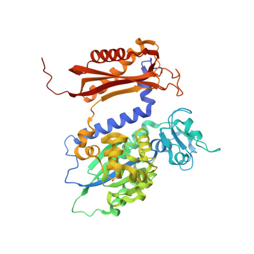
6X77

Rev1 R518A Ternary Complex with dCTP and Ca2+


Experimental Data Snapshot

  • Method: X-RAY DIFFRACTION
  • Resolution: 1.64 Å
  • R-Value Free: 0.221 
  • R-Value Work: 0.180 
  • R-Value Observed: 0.182 

Starting Model: experimental
View more details

wwPDB Validation   3D Report Full Report


Ligand Structure Quality Assessment 


This is version 1.2 of the entry. See complete history


Literature

Visualizing Rev1 catalyze protein-template DNA synthesis.

Weaver, T.M.Cortez, L.M.Khoang, T.H.Washington, M.T.Agarwal, P.K.Freudenthal, B.D.

(2020) Proc Natl Acad Sci U S A 117: 25494-25504

  • DOI: https://doi.org/10.1073/pnas.2010484117
  • Primary Citation of Related Structures:  
    6X6Z, 6X70, 6X71, 6X72, 6X73, 6X74, 6X75, 6X76, 6X77

  • PubMed Abstract: 

    During DNA replication, replicative DNA polymerases may encounter DNA lesions, which can stall replication forks. One way to prevent replication fork stalling is through the recruitment of specialized translesion synthesis (TLS) polymerases that have evolved to incorporate nucleotides opposite DNA lesions. Rev1 is a specialized TLS polymerase that bypasses abasic sites, as well as minor-groove and exocyclic guanine adducts. Lesion bypass is accomplished using a unique protein-template mechanism in which the templating base is evicted from the DNA helix and the incoming dCTP hydrogen bonds with an arginine side chain of Rev1. To understand the protein-template mechanism at an atomic level, we employed a combination of time-lapse X-ray crystallography, molecular dynamics simulations, and DNA enzymology on the Saccharomyces cerevisiae Rev1 protein. We find that Rev1 evicts the templating base from the DNA helix prior to binding the incoming nucleotide. Binding the incoming nucleotide changes the conformation of the DNA substrate to orient it for nucleotidyl transfer, although this is not coupled to large structural changes in Rev1 like those observed with other DNA polymerases. Moreover, we found that following nucleotide incorporation, Rev1 converts the pyrophosphate product to two monophosphates, which drives the reaction in the forward direction and prevents pyrophosphorolysis. Following nucleotide incorporation, the hydrogen bonds between the incorporated nucleotide and the arginine side chain are broken, but the templating base remains extrahelical. These postcatalytic changes prevent potentially mutagenic processive synthesis by Rev1 and facilitate dissociation of the DNA product from the enzyme.


  • Organizational Affiliation

    Department of Biochemistry and Molecular Biology, University of Kansas Medical Center, Kansas City, KS 66160.


Macromolecules

Find similar proteins by:  (by identity cutoff)  |  3D Structure
Entity ID: 3
MoleculeChains Sequence LengthOrganismDetailsImage
DNA repair protein REV1C [auth A]442Saccharomyces cerevisiaeMutation(s): 1 
Gene Names: REV1YOR346WO6339
EC: 2.7.7
UniProt
Find proteins for P12689 (Saccharomyces cerevisiae (strain ATCC 204508 / S288c))
Explore P12689 
Go to UniProtKB:  P12689
Entity Groups  
Sequence Clusters30% Identity50% Identity70% Identity90% Identity95% Identity100% Identity
UniProt GroupP12689
Sequence Annotations
Expand
  • Reference Sequence

Find similar nucleic acids by:  Sequence   |   3D Structure  

Entity ID: 1
MoleculeChains LengthOrganismImage
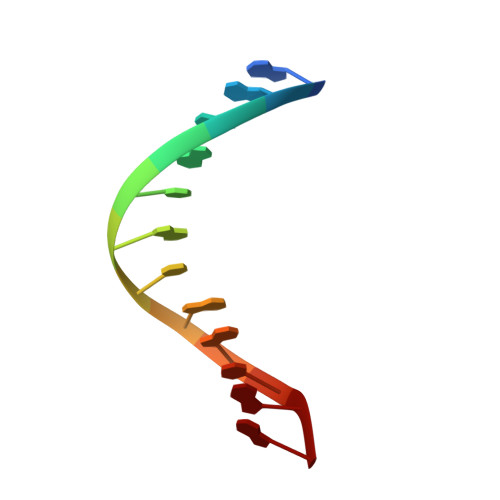
DNA (5'-D(P*GP*GP*GP*GP*TP*GP*TP*GP*GP*TP*AP*G)-3')A [auth P]12synthetic construct
Sequence Annotations
Expand
  • Reference Sequence

Find similar nucleic acids by:  Sequence   |   3D Structure  

Entity ID: 2
MoleculeChains LengthOrganismImage
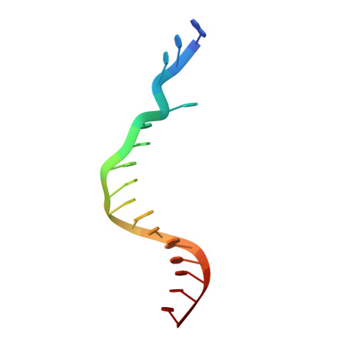
DNA (5'-D(P*AP*TP*CP*GP*CP*TP*AP*CP*CP*AP*CP*AP*CP*CP*CP*C)-3')B [auth T]16synthetic construct
Sequence Annotations
Expand
  • Reference Sequence
Experimental Data & Validation

Experimental Data

  • Method: X-RAY DIFFRACTION
  • Resolution: 1.64 Å
  • R-Value Free: 0.221 
  • R-Value Work: 0.180 
  • R-Value Observed: 0.182 
  • Space Group: P 21 21 21
Unit Cell:
Length ( Å )Angle ( ˚ )
a = 62.258α = 90
b = 73.771β = 90
c = 116.811γ = 90
Software Package:
Software NamePurpose
PHENIXrefinement
HKL-3000data reduction
HKL-3000data scaling
PHASERphasing

Structure Validation

View Full Validation Report


Ligand Structure Quality Assessment 


Entry History & Funding Information

Deposition Data


Funding OrganizationLocationGrant Number
National Institutes of Health/National Institute of General Medical Sciences (NIH/NIGMS)United States--

Revision History  (Full details and data files)

  • Version 1.0: 2020-09-30
    Type: Initial release
  • Version 1.1: 2020-10-21
    Changes: Database references
  • Version 1.2: 2023-10-18
    Changes: Data collection, Database references, Refinement description