6Y0Q

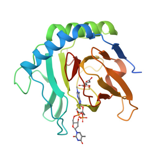
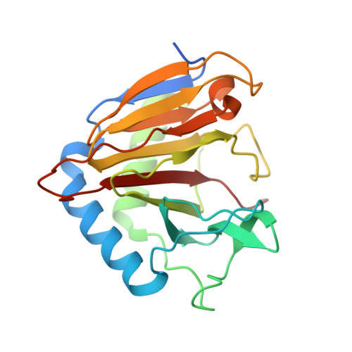
Alpha-ketoglutarate-dependent dioxygenase AlkB in complex with Fe, AKG and methylated DNA under anaerobic environment using FT-SSX methods


Experimental Data Snapshot

  • Method: X-RAY DIFFRACTION
  • Resolution: 1.75 Å
  • R-Value Free: 0.191 
  • R-Value Work: 0.162 
  • R-Value Observed: 0.165 

Starting Model: experimental
View more details

wwPDB Validation   3D Report Full Report


Ligand Structure Quality Assessment 


This is version 1.2 of the entry. See complete history


Literature

Anaerobic fixed-target serial crystallography.

Rabe, P.Beale, J.H.Butryn, A.Aller, P.Dirr, A.Lang, P.A.Axford, D.N.Carr, S.B.Leissing, T.M.McDonough, M.A.Davy, B.Ebrahim, A.Orlans, J.Storm, S.L.S.Orville, A.M.Schofield, C.J.Owen, R.L.

(2020) IUCrJ 7: 901-912

  • DOI: https://doi.org/10.1107/S2052252520010374
  • Primary Citation of Related Structures:  
    6Y0N, 6Y0O, 6Y0Q, 6Y12, 6YPV

  • PubMed Abstract: 

    Cryogenic X-ray diffraction is a powerful tool for crystallographic studies on enzymes including oxygenases and oxidases. Amongst the benefits that cryo-conditions (usually employing a nitro-gen cryo-stream at 100 K) enable, is data collection of di-oxy-gen-sensitive samples. Although not strictly anaerobic, at low temperatures the vitreous ice conditions severely restrict O 2 diffusion into and/or through the protein crystal. Cryo-conditions limit chemical reactivity, including reactions that require significant conformational changes. By contrast, data collection at room temperature imposes fewer restrictions on diffusion and reactivity; room-temperature serial methods are thus becoming common at synchrotrons and XFELs. However, maintaining an anaerobic environment for di-oxy-gen-dependent enzymes has not been explored for serial room-temperature data collection at synchrotron light sources. This work describes a methodology that employs an adaptation of the 'sheet-on-sheet' sample mount, which is suitable for the low-dose room-temperature data collection of anaerobic samples at synchrotron light sources. The method is characterized by easy sample preparation in an anaerobic glovebox, gentle handling of crystals, low sample consumption and preservation of a localized anaerobic environment over the timescale of the experiment (<5 min). The utility of the method is highlighted by studies with three X-ray-radiation-sensitive Fe(II)-containing model enzymes: the 2-oxoglutarate-dependent l-arginine hy-droxy-lase VioC and the DNA repair enzyme AlkB, as well as the oxidase isopenicillin N synthase (IPNS), which is involved in the biosynthesis of all penicillin and cephalosporin antibiotics.


  • Organizational Affiliation

    Chemistry Research Laboratory, University of Oxford, 12 Mansfield Road, Oxford OX1 3TA, United Kingdom.


Macromolecules
Find similar proteins by:  (by identity cutoff)  |  3D Structure
Entity ID: 1
MoleculeChains Sequence LengthOrganismDetailsImage
Alpha-ketoglutarate-dependent dioxygenase AlkB216Escherichia coli K-12Mutation(s): 0 
Gene Names: alkBaidDb2212JW2200
EC: 1.14.11.33
UniProt
Find proteins for P05050 (Escherichia coli (strain K12))
Explore P05050 
Go to UniProtKB:  P05050
Entity Groups  
Sequence Clusters30% Identity50% Identity70% Identity90% Identity95% Identity100% Identity
UniProt GroupP05050
Sequence Annotations
Expand
  • Reference Sequence
Small Molecules
Ligands 3 Unique
IDChains Name / Formula / InChI Key2D Diagram3D Interactions
O5W (Subject of Investigation/LOI)
Query on O5W

Download Ideal Coordinates CCD File 
D [auth A][(3~{S},5~{R})-5-(6-azanyl-1-methyl-purin-9-yl)-2-(phosphonooxymethyl)oxolan-3-yl] [(2~{R},3~{S},5~{R})-5-[5-methyl-2,4-bis(oxidanylidene)pyrimidin-1-yl]-3-oxidanyl-oxolan-2-yl]methyl hydrogen phosphate
C21 H28 N7 O13 P2
AESRSXSUEFZXFW-KPRKPIBOSA-O
AKG
Query on AKG

Download Ideal Coordinates CCD File 
B [auth A]2-OXOGLUTARIC ACID
C5 H6 O5
KPGXRSRHYNQIFN-UHFFFAOYSA-N
FE
Query on FE

Download Ideal Coordinates CCD File 
C [auth A]FE (III) ION
Fe
VTLYFUHAOXGGBS-UHFFFAOYSA-N
Experimental Data & Validation

Experimental Data

  • Method: X-RAY DIFFRACTION
  • Resolution: 1.75 Å
  • R-Value Free: 0.191 
  • R-Value Work: 0.162 
  • R-Value Observed: 0.165 
  • Space Group: P 1
Unit Cell:
Length ( Å )Angle ( ˚ )
a = 36.399α = 78.99
b = 39.078β = 78.04
c = 40.84γ = 66.88
Software Package:
Software NamePurpose
PHENIXrefinement
GDAdata collection
DIALSdata reduction
DIALSdata scaling
PHASERphasing

Structure Validation

View Full Validation Report


Ligand Structure Quality Assessment 


Entry History & Funding Information

Deposition Data


Funding OrganizationLocationGrant Number
Wellcome TrustUnited Kingdom106244/Z/14/Z
Medical Research Council (United Kingdom)United KingdomMR/N002679/1
Biotechnology and Biological Sciences Research CouncilUnited KingdomBB/S50676X/1

Revision History  (Full details and data files)

  • Version 1.0: 2020-09-09
    Type: Initial release
  • Version 1.1: 2020-09-30
    Changes: Database references
  • Version 1.2: 2024-01-24
    Changes: Data collection, Database references, Refinement description