6ZI2

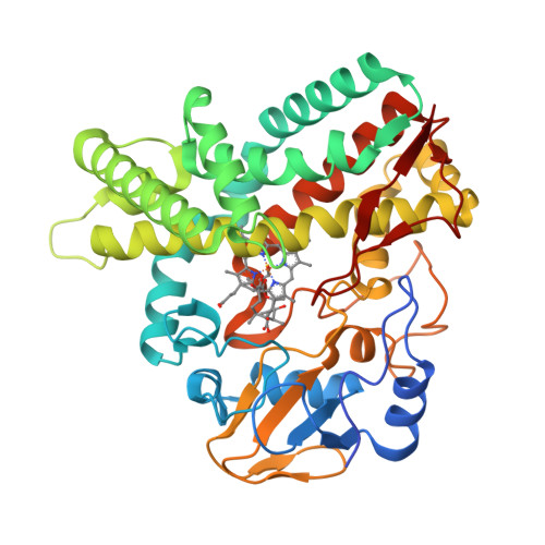
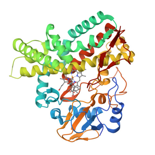
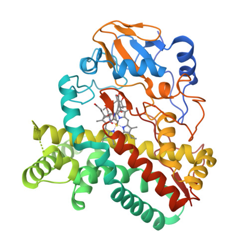
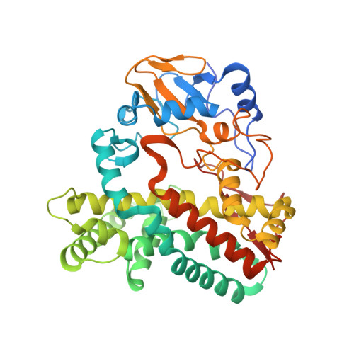
OleP-oleandolide(DEO) in low salt crystallization conditions


Experimental Data Snapshot

  • Method: X-RAY DIFFRACTION
  • Resolution: 2.93 Å
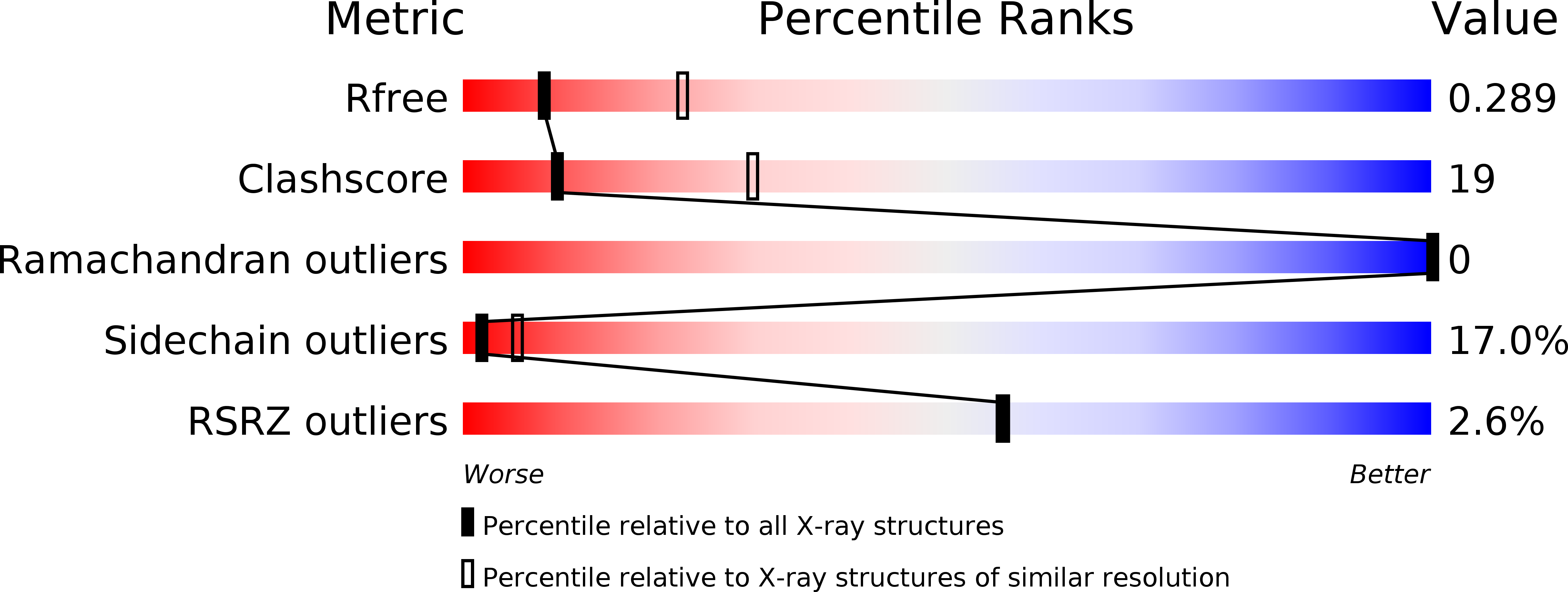
  • R-Value Free: 0.291 
  • R-Value Work: 0.239 
  • R-Value Observed: 0.242 

Starting Model: experimental
View more details

wwPDB Validation   3D Report Full Report


Ligand Structure Quality Assessment 


This is version 1.1 of the entry. See complete history


Literature

Dissecting the Cytochrome P450 OleP Substrate Specificity: Evidence for a Preferential Substrate.

Parisi, G.Freda, I.Exertier, C.Cecchetti, C.Gugole, E.Cerutti, G.D'Auria, L.Macone, A.Vallone, B.Savino, C.Montemiglio, L.C.

(2020) Biomolecules 10

  • DOI: https://doi.org/10.3390/biom10101411
  • Primary Citation of Related Structures:  
    6ZHZ, 6ZI2, 6ZI3, 6ZI7

  • PubMed Abstract: 

    The cytochrome P450 OleP catalyzes the epoxidation of aliphatic carbons on both the aglycone 8.8a-deoxyoleandolide (DEO) and the monoglycosylated L-olivosyl-8.8a-deoxyoleandolide (L-O-DEO) intermediates of oleandomycin biosynthesis. We investigated the substrate versatility of the enzyme. X-ray and equilibrium binding data show that the aglycone DEO loosely fits the OleP active site, triggering the closure that prepares it for catalysis only on a minor population of enzyme. The open-to-closed state transition allows solvent molecules to accumulate in a cavity that forms upon closure, mediating protein-substrate interactions. In silico docking of the monoglycosylated L-O-DEO in the closed OleP-DEO structure shows that the L-olivosyl moiety can be hosted in the same cavity, replacing solvent molecules and directly contacting structural elements involved in the transition. X-ray structures of aglycone-bound OleP in the presence of L-rhamnose confirm the cavity as a potential site for sugar binding. All considered, we propose L-O-DEO as the optimal substrate of OleP, the L-olivosyl moiety possibly representing the molecular wedge that triggers a more efficient structural response upon substrate binding, favoring and stabilizing the enzyme closure before catalysis. OleP substrate versatility is supported by structural solvent molecules that compensate for the absence of a glycosyl unit when the aglycone is bound.


  • Organizational Affiliation

    Istituto Pasteur-Fondazione Cenci Bolognetti and Department of Biochemical Sciences "Alessandro Rossi Fanelli", Sapienza, University of Rome, P. le Aldo Moro, 5, 00185 Rome, Italy.


Macromolecules
Find similar proteins by:  (by identity cutoff)  |  3D Structure
Entity ID: 1
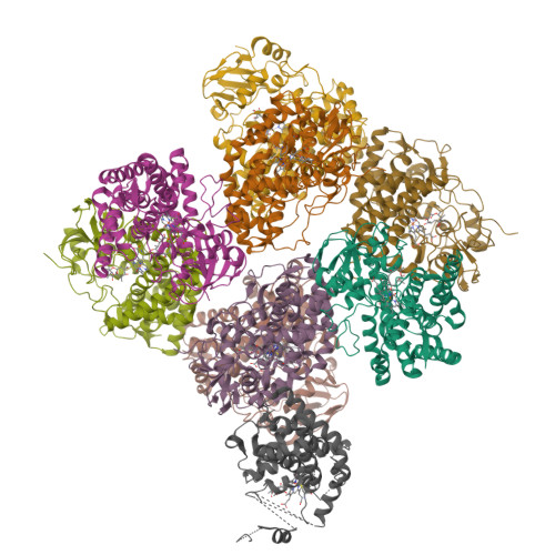
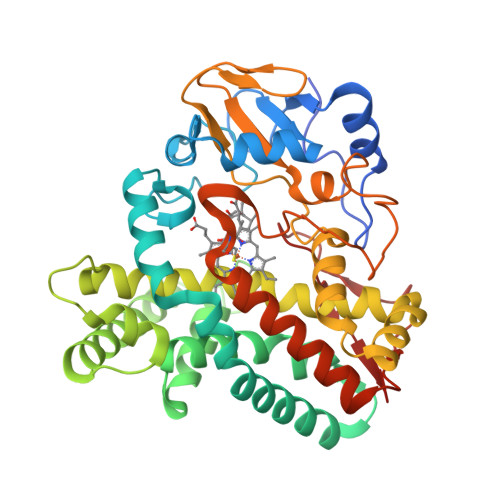
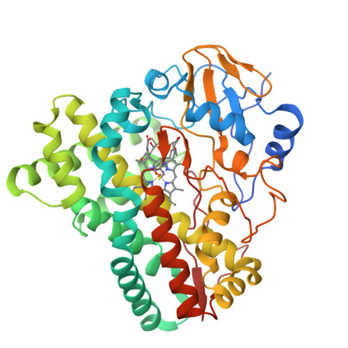
MoleculeChains Sequence LengthOrganismDetailsImage
Cytochrome P-450
A, B, C, D, E
A, B, C, D, E, F, G, H, I
407Streptomyces antibioticusMutation(s): 0 
Gene Names: oleP
UniProt
Find proteins for Q59819 (Streptomyces antibioticus)
Explore Q59819 
Go to UniProtKB:  Q59819
Entity Groups  
Sequence Clusters30% Identity50% Identity70% Identity90% Identity95% Identity100% Identity
UniProt GroupQ59819
Sequence Annotations
Expand
  • Reference Sequence
Small Molecules
Ligands 2 Unique
IDChains Name / Formula / InChI Key2D Diagram3D Interactions
HEM
Query on HEM

Download Ideal Coordinates CCD File 
J [auth A]
L [auth B]
N [auth C]
P [auth D]
R [auth E]
J [auth A],
L [auth B],
N [auth C],
P [auth D],
R [auth E],
T [auth F],
V [auth G],
X [auth H],
Z [auth I]
PROTOPORPHYRIN IX CONTAINING FE
C34 H32 Fe N4 O4
KABFMIBPWCXCRK-RGGAHWMASA-L
QR8 (Subject of Investigation/LOI)
Query on QR8

Download Ideal Coordinates CCD File 
AA [auth I]
K [auth A]
M [auth B]
O [auth C]
Q [auth D]
AA [auth I],
K [auth A],
M [auth B],
O [auth C],
Q [auth D],
S [auth E],
U [auth F],
W [auth G],
Y [auth H]
(3~{R},4~{S},5~{R},6~{S},7~{S},9~{S},11~{R},12~{S},13~{R},14~{R})-3,5,7,9,11,13,14-heptamethyl-4,6,12-tris(oxidanyl)-1-oxacyclotetradecane-2,10-dione
C20 H36 O6
OLPAVFVRSHWADO-ODNSKKOKSA-N
Experimental Data & Validation

Experimental Data

  • Method: X-RAY DIFFRACTION
  • Resolution: 2.93 Å
  • R-Value Free: 0.291 
  • R-Value Work: 0.239 
  • R-Value Observed: 0.242 
  • Space Group: P 1
Unit Cell:
Length ( Å )Angle ( ˚ )
a = 112.066α = 104.43
b = 116.642β = 104.25
c = 125.17γ = 113.91
Software Package:
Software NamePurpose
REFMACrefinement
XDSdata reduction
XDSdata scaling
MOLREPphasing

Structure Validation

View Full Validation Report


Ligand Structure Quality Assessment 


Entry History & Funding Information

Deposition Data


Funding OrganizationLocationGrant Number
Pasteur InstituteItaly--

Revision History  (Full details and data files)

  • Version 1.0: 2020-10-21
    Type: Initial release
  • Version 1.1: 2024-01-31
    Changes: Data collection, Database references, Refinement description