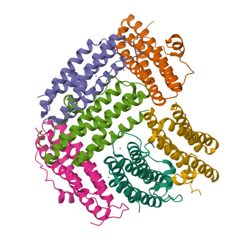
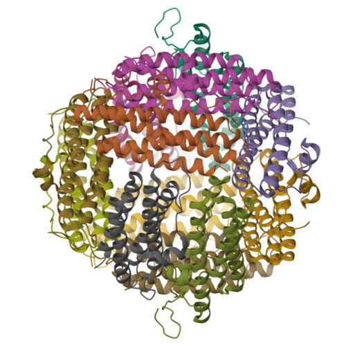
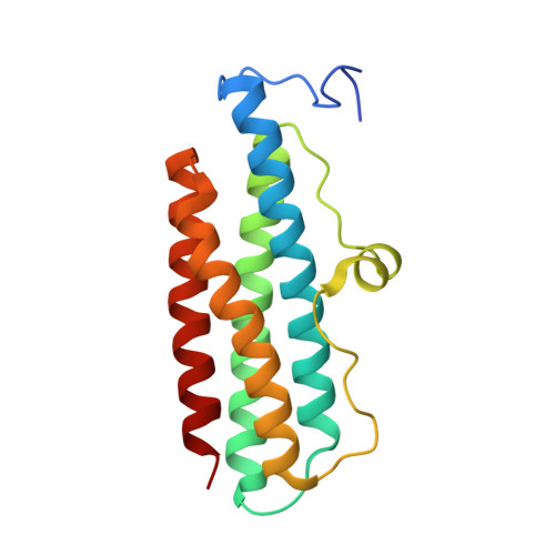
SIMBAD: a sequence-independent molecular-replacement pipeline.
Simpkin, A.J., Simkovic, F., Thomas, J.M.H., Savko, M., Lebedev, A., Uski, V., Ballard, C., Wojdyr, M., Wu, R., Sanishvili, R., Xu, Y., Lisa, M.N., Buschiazzo, A., Shepard, W., Rigden, D.J., Keegan, R.M.(2018) Acta Crystallogr D Struct Biol 74: 595-605
- PubMed: 29968670
- DOI: https://doi.org/10.1107/S2059798318005752
- Primary Citation of Related Structures:
6B0D, 6B6M, 6BY0 - PubMed Abstract:
The conventional approach to finding structurally similar search models for use in molecular replacement (MR) is to use the sequence of the target to search against those of a set of known structures. Sequence similarity often correlates with structure similarity. Given sufficient similarity, a known structure correctly positioned in the target cell by the MR process can provide an approximation to the unknown phases of the target. An alternative approach to identifying homologous structures suitable for MR is to exploit the measured data directly, comparing the lattice parameters or the experimentally derived structure-factor amplitudes with those of known structures. Here, SIMBAD, a new sequence-independent MR pipeline which implements these approaches, is presented. SIMBAD can identify cases of contaminant crystallization and other mishaps such as mistaken identity (swapped crystallization trays), as well as solving unsequenced targets and providing a brute-force approach where sequence-dependent search-model identification may be nontrivial, for example because of conformational diversity among identifiable homologues. The program implements a three-step pipeline to efficiently identify a suitable search model in a database of known structures. The first step performs a lattice-parameter search against the entire Protein Data Bank (PDB), rapidly determining whether or not a homologue exists in the same crystal form. The second step is designed to screen the target data for the presence of a crystallized contaminant, a not uncommon occurrence in macromolecular crystallography. Solving structures with MR in such cases can remain problematic for many years, since the search models, which are assumed to be similar to the structure of interest, are not necessarily related to the structures that have actually crystallized. To cater for this eventuality, SIMBAD rapidly screens the data against a database of known contaminant structures. Where the first two steps fail to yield a solution, a final step in SIMBAD can be invoked to perform a brute-force search of a nonredundant PDB database provided by the MoRDa MR software. Through early-access usage of SIMBAD, this approach has solved novel cases that have otherwise proved difficult to solve.
Organizational Affiliation:
Institute of Integrative Biology, University of Liverpool, Liverpool L69 7ZB, England.


