6BW5

Human GPT (DPAGT1) in complex with tunicamycin


Experimental Data Snapshot

  • Method: X-RAY DIFFRACTION
  • Resolution: 3.10 Å
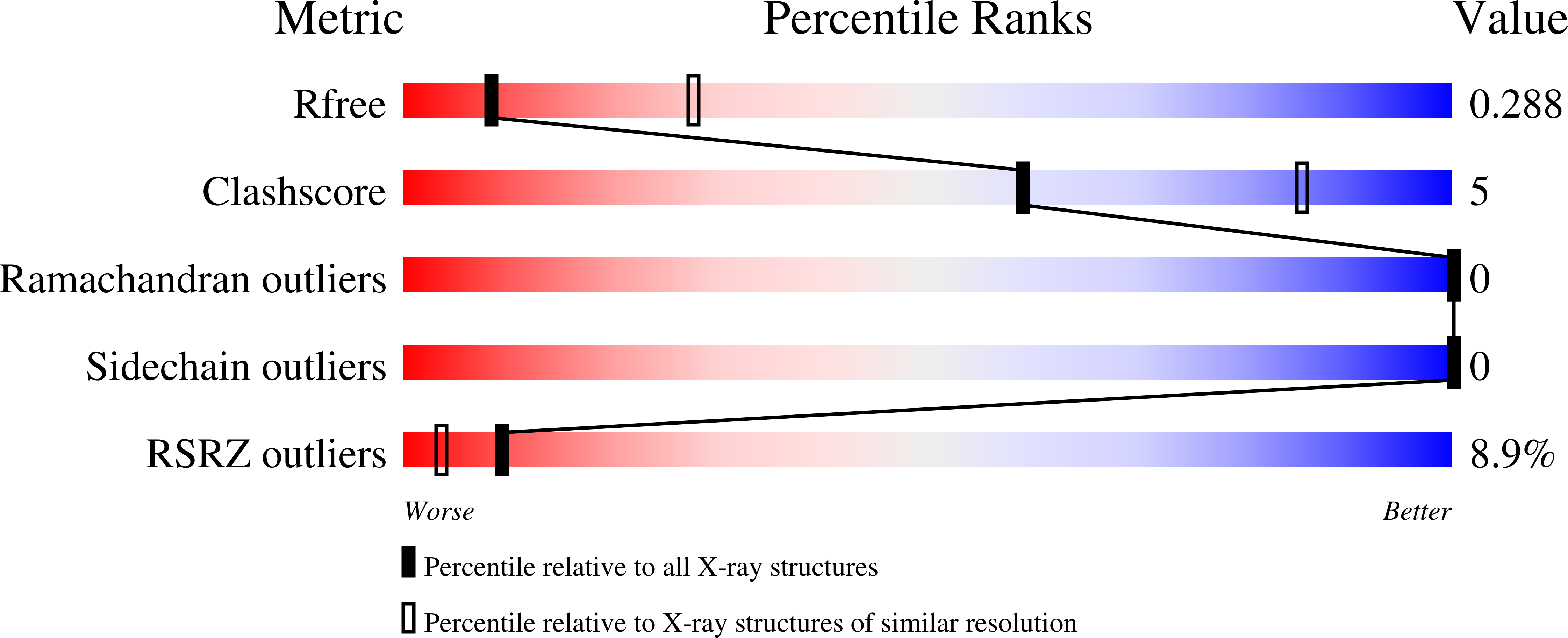
  • R-Value Free: 0.288 
  • R-Value Work: 0.256 
  • R-Value Observed: 0.258 

Starting Model: experimental
View more details

wwPDB Validation   3D Report Full Report


Ligand Structure Quality Assessment 


This is version 1.6 of the entry. See complete history


Literature

GlcNAc-1-P-transferase-tunicamycin complex structure reveals basis for inhibition of N-glycosylation.

Yoo, J.Mashalidis, E.H.Kuk, A.C.Y.Yamamoto, K.Kaeser, B.Ichikawa, S.Lee, S.Y.

(2018) Nat Struct Mol Biol 25: 217-224

  • DOI: https://doi.org/10.1038/s41594-018-0031-y
  • Primary Citation of Related Structures:  
    6BW5, 6BW6

  • PubMed Abstract: 

    N-linked glycosylation is a predominant post-translational modification of protein in eukaryotes, and its dysregulation is the etiology of several human disorders. The enzyme UDP-N-acetylglucosamine:dolichyl-phosphate N-acetylglucosaminephosphotransferase (GlcNAc-1-P-transferase or GPT) catalyzes the first and committed step of N-linked glycosylation in the endoplasmic reticulum membrane, and it is the target of the natural product tunicamycin. Tunicamycin has potent antibacterial activity, inhibiting the bacterial cell wall synthesis enzyme MraY, but its usefulness as an antibiotic is limited by off-target inhibition of human GPT. Our understanding of how tunicamycin inhibits N-linked glycosylation and efforts to selectively target MraY are hampered by a lack of structural information. Here we present crystal structures of human GPT in complex with tunicamycin. Structural and functional analyses reveal the difference between GPT and MraY in their mechanisms of inhibition by tunicamycin. We demonstrate that this difference could be exploited to design MraY-specific inhibitors as potential antibiotics.


  • Organizational Affiliation

    Department of Biochemistry, Duke University Medical Center, Durham, NC, USA.


Macromolecules
Find similar proteins by:  (by identity cutoff)  |  3D Structure
Entity ID: 1
MoleculeChains Sequence LengthOrganismDetailsImage
UDP-N-acetylglucosamine--dolichyl-phosphate N-acetylglucosaminephosphotransferase
A, B, C, D
417Homo sapiensMutation(s): 0 
Gene Names: DPAGT1DPAGT2
EC: 2.7.8.15
Membrane Entity: Yes 
UniProt & NIH Common Fund Data Resources
Find proteins for Q9H3H5 (Homo sapiens)
Explore Q9H3H5 
Go to UniProtKB:  Q9H3H5
PHAROS:  Q9H3H5
GTEx:  ENSG00000172269 
Entity Groups  
Sequence Clusters30% Identity50% Identity70% Identity90% Identity95% Identity100% Identity
UniProt GroupQ9H3H5
Sequence Annotations
Expand
  • Reference Sequence
Small Molecules
Ligands 2 Unique
IDChains Name / Formula / InChI Key2D Diagram3D Interactions
TUM (Subject of Investigation/LOI)
Query on TUM

Download Ideal Coordinates CCD File 
E [auth A],
G [auth B],
I [auth C],
K [auth D]
Tunicamycin
C37 H60 N4 O16
YJQCOFNZVFGCAF-DCSBULBVSA-N
PGW
Query on PGW

Download Ideal Coordinates CCD File 
F [auth A],
H [auth B],
J [auth C],
L [auth D]
(1R)-2-{[(S)-{[(2S)-2,3-dihydroxypropyl]oxy}(hydroxy)phosphoryl]oxy}-1-[(hexadecanoyloxy)methyl]ethyl (9Z)-octadec-9-enoate
C40 H77 O10 P
PAZGBAOHGQRCBP-HGWHEPCSSA-N
Experimental Data & Validation

Experimental Data

  • Method: X-RAY DIFFRACTION
  • Resolution: 3.10 Å
  • R-Value Free: 0.288 
  • R-Value Work: 0.256 
  • R-Value Observed: 0.258 
  • Space Group: C 1 2 1
Unit Cell:
Length ( Å )Angle ( ˚ )
a = 212.272α = 90
b = 105.512β = 103.53
c = 149.389γ = 90
Software Package:
Software NamePurpose
PHENIXrefinement
XDSdata reduction
Aimlessdata scaling
PHASERphasing

Structure Validation

View Full Validation Report


Ligand Structure Quality Assessment 


Entry History & Funding Information

Deposition Data


Funding OrganizationLocationGrant Number
National Institutes of Health/National Institute of General Medical Sciences (NIH/NIGMS)United StatesR01GM120594
National Institutes of Health/National Institute of Neurological Disorders and Stroke (NIH/NINDS)United StatesR35NS097241

Revision History  (Full details and data files)

  • Version 1.0: 2018-02-21
    Type: Initial release
  • Version 1.1: 2018-02-28
    Changes: Author supporting evidence
  • Version 1.2: 2018-03-14
    Changes: Database references
  • Version 1.3: 2018-03-21
    Changes: Database references
  • Version 1.4: 2019-12-18
    Changes: Author supporting evidence
  • Version 1.5: 2023-10-04
    Changes: Data collection, Database references, Refinement description
  • Version 1.6: 2024-11-20
    Changes: Structure summary