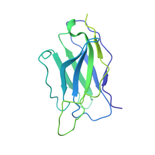
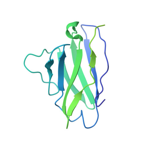
6CHB

Crystal structure of a natively-glycosylated BG505 SOSIP.664 HIV-1 Envelope Trimer in complex with the broadly-neutralizing antibodies BG18 and IOMA


Experimental Data Snapshot

  • Method: X-RAY DIFFRACTION
  • Resolution: 6.80 Å
  • R-Value Free: 0.417 
  • R-Value Work: 0.318 
  • R-Value Observed: 0.323 

Starting Models: experimental
View more details

wwPDB Validation   3D Report Full Report


This is version 1.3 of the entry. See complete history


Literature

Structural characterization of a highly-potent V3-glycan broadly neutralizing antibody bound to natively-glycosylated HIV-1 envelope.

Barnes, C.O.Gristick, H.B.Freund, N.T.Escolano, A.Lyubimov, A.Y.Hartweger, H.West, A.P.Cohen, A.E.Nussenzweig, M.C.Bjorkman, P.J.

(2018) Nat Commun 9: 1251-1251

  • DOI: https://doi.org/10.1038/s41467-018-03632-y
  • Primary Citation of Related Structures:  
    6CH7, 6CH8, 6CH9, 6CHB

  • PubMed Abstract: 

    Broadly neutralizing antibodies (bNAbs) isolated from HIV-1-infected individuals inform HIV-1 vaccine design efforts. Developing bNAbs with increased efficacy requires understanding how antibodies interact with the native oligomannose and complex-type N-glycan shield that hides most protein epitopes on HIV-1 envelope (Env). Here we present crystal structures, including a 3.8-Å X-ray free electron laser dataset, of natively glycosylated Env trimers complexed with BG18, the most potent V3/N332 gp120 glycan-targeting bNAb reported to date. Our structures show conserved contacts mediated by common D gene-encoded residues with the N332 gp120 glycan and the gp120 GDIR peptide motif, but a distinct Env-binding orientation relative to PGT121/10-1074 bNAbs. BG18's binding orientation provides additional contacts with N392 gp120 and N386 gp120 glycans near the V3-loop base and engages protein components of the V1-loop. The BG18-natively-glycosylated Env structures facilitate understanding of bNAb-glycan interactions critical for using V3/N332 gp120 bNAbs therapeutically and targeting their epitope for immunogen design.


  • Organizational Affiliation

    Division of Biology and Biological Engineering, California Institute of Technology, Pasadena, CA, 91125, USA.


Macromolecules
Find similar proteins by:  (by identity cutoff)  |  3D Structure
Entity ID: 1
MoleculeChains Sequence LengthOrganismDetailsImage
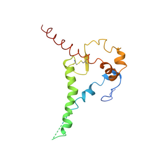
Envelope glycoprotein gp41A [auth B],
G [auth A],
M [auth C]
153Human immunodeficiency virus 1Mutation(s): 1 
Gene Names: env
UniProt
Find proteins for Q2N0S5 (Human immunodeficiency virus 1)
Explore Q2N0S5 
Go to UniProtKB:  Q2N0S5
Entity Groups  
Sequence Clusters30% Identity50% Identity70% Identity90% Identity95% Identity100% Identity
UniProt GroupQ2N0S5
Sequence Annotations
Expand
  • Reference Sequence
Find similar proteins by:  (by identity cutoff)  |  3D Structure
Entity ID: 2
MoleculeChains Sequence LengthOrganismDetailsImage
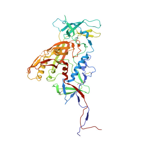
Envelope glycoprotein gp120B [auth G],
H [auth F],
N [auth H]
479Human immunodeficiency virus 1Mutation(s): 1 
Gene Names: env
UniProt
Find proteins for Q2N0S6 (Human immunodeficiency virus 1)
Explore Q2N0S6 
Go to UniProtKB:  Q2N0S6
Entity Groups  
Sequence Clusters30% Identity50% Identity70% Identity90% Identity95% Identity100% Identity
UniProt GroupQ2N0S6
Sequence Annotations
Expand
  • Reference Sequence
Find similar proteins by:  (by identity cutoff)  |  3D Structure
Entity ID: 3
MoleculeChains Sequence LengthOrganismDetailsImage
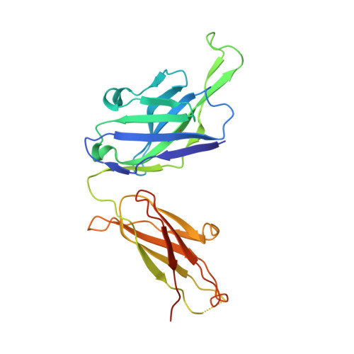
BG18 Heavy ChainC [auth J],
K [auth I],
Q
241Homo sapiensMutation(s): 0 
Entity Groups  
Sequence Clusters30% Identity50% Identity70% Identity90% Identity95% Identity100% Identity
Sequence Annotations
Expand
  • Reference Sequence
Find similar proteins by:  (by identity cutoff)  |  3D Structure
Entity ID: 4
MoleculeChains Sequence LengthOrganismDetailsImage
BG18 Light ChainD [auth K],
L,
R
215Homo sapiensMutation(s): 0 
Entity Groups  
Sequence Clusters30% Identity50% Identity70% Identity90% Identity95% Identity100% Identity
Sequence Annotations
Expand
  • Reference Sequence
Find similar proteins by:  (by identity cutoff)  |  3D Structure
Entity ID: 5
MoleculeChains Sequence LengthOrganismDetailsImage
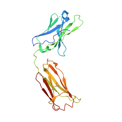
IOMA Heavy ChainE [auth D],
I [auth M],
O
232Homo sapiensMutation(s): 0 
Entity Groups  
Sequence Clusters30% Identity50% Identity70% Identity90% Identity95% Identity100% Identity
Sequence Annotations
Expand
  • Reference Sequence
Find similar proteins by:  (by identity cutoff)  |  3D Structure
Entity ID: 6
MoleculeChains Sequence LengthOrganismDetailsImage
IOMA Light ChainF [auth E],
J [auth N],
P
214Homo sapiensMutation(s): 0 
Entity Groups  
Sequence Clusters30% Identity50% Identity70% Identity90% Identity95% Identity100% Identity
Sequence Annotations
Expand
  • Reference Sequence
Experimental Data & Validation

Experimental Data

  • Method: X-RAY DIFFRACTION
  • Resolution: 6.80 Å
  • R-Value Free: 0.417 
  • R-Value Work: 0.318 
  • R-Value Observed: 0.323 
  • Space Group: P 43 21 2
Unit Cell:
Length ( Å )Angle ( ˚ )
a = 176.752α = 90
b = 176.752β = 90
c = 458.039γ = 90
Software Package:
Software NamePurpose
PHENIXrefinement
Aimlessdata scaling
PDB_EXTRACTdata extraction
XDSdata reduction
PHASERphasing

Structure Validation

View Full Validation Report


Entry History & Funding Information

Deposition Data


Funding OrganizationLocationGrant Number
National Institutes of Health/National Institute Of Allergy and Infectious Diseases (NIH/NIAID)United StatesP01 AI100148
Bill & Melinda Gates FoundationUnited StatesOPP1124068
National Institutes of Health/National Institute of General Medical Sciences (NIH/NIGMS)United StatesP41GM103393

Revision History  (Full details and data files)

  • Version 1.0: 2018-05-02
    Type: Initial release
  • Version 1.1: 2019-02-20
    Changes: Author supporting evidence, Data collection
  • Version 1.2: 2019-12-18
    Changes: Author supporting evidence
  • Version 1.3: 2023-10-04
    Changes: Data collection, Database references, Refinement description