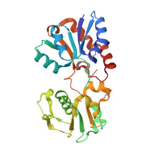
6CVA

Crystal structure of the N. meningitides methionine-binding protein in its substrate-free conformation


Experimental Data Snapshot

  • Method: X-RAY DIFFRACTION
  • Resolution: 1.56 Å
  • R-Value Free: 0.220 
  • R-Value Work: 0.188 
  • R-Value Observed: 0.190 

wwPDB Validation   3D Report Full Report


This is version 1.4 of the entry. See complete history


Literature

Structures of the Neisseria meningitides methionine-binding protein MetQ in substrate-free form and bound to l- and d-methionine isomers.

Nguyen, P.T.Lai, J.Y.Kaiser, J.T.Rees, D.C.

(2019) Protein Sci 28: 1750-1757

  • DOI: https://doi.org/10.1002/pro.3694
  • Primary Citation of Related Structures:  
    6CVA, 6DZX, 6OJA

  • PubMed Abstract: 

    The bacterial periplasmic methionine-binding protein MetQ is involved in the import of methionine by the cognate MetNI methionine ATP binding cassette (ABC) transporter. The MetNIQ system is one of the few members of the ABC importer family that has been structurally characterized in multiple conformational states. Critical missing elements in the structural analysis of MetNIQ are the structure of the substrate-free form of MetQ, and detailing how MetQ binds multiple methionine derivatives, including both l- and d-methionine isomers. In this study, we report the structures of the Neisseria meningitides MetQ in substrate-free form and in complexes with l-methionine and with d-methionine, along with the associated binding constants determined by isothermal titration calorimetry. Structures of the substrate-free (N238A) and substrate-bound N. meningitides MetQ are related by a "Venus-fly trap" hinge-type movement of the two domains accompanying methionine binding and dissociation. l- and d-methionine bind to the same site on MetQ, and this study emphasizes the important role of asparagine 238 in ligand binding and affinity. A thermodynamic analysis demonstrates that ligand-free MetQ associates with the ATP-bound form of MetNI ∼40 times more tightly than does liganded MetQ, consistent with the necessity of dissociating methionine from MetQ for transport to occur.


  • Organizational Affiliation

    Division of Chemistry and Chemical Engineering, California Institute of Technology, Pasadena, California.


Macromolecules
Find similar proteins by:  (by identity cutoff)  |  3D Structure
Entity ID: 1
MoleculeChains Sequence LengthOrganismDetailsImage
Lipoprotein241Neisseria meningitidisMutation(s): 0 
Gene Names: 
UniProt
Find proteins for Q9JPG4 (Neisseria meningitidis)
Explore Q9JPG4 
Go to UniProtKB:  Q9JPG4
Entity Groups  
Sequence Clusters30% Identity50% Identity70% Identity90% Identity95% Identity100% Identity
UniProt GroupQ9JPG4
Sequence Annotations
Expand
  • Reference Sequence
Small Molecules
Modified Residues  1 Unique
IDChains TypeFormula2D DiagramParent
MSE
Query on MSE
A
L-PEPTIDE LINKINGC5 H11 N O2 SeMET
Experimental Data & Validation

Experimental Data

  • Method: X-RAY DIFFRACTION
  • Resolution: 1.56 Å
  • R-Value Free: 0.220 
  • R-Value Work: 0.188 
  • R-Value Observed: 0.190 
  • Space Group: P 21 21 2
Unit Cell:
Length ( Å )Angle ( ˚ )
a = 52.564α = 90
b = 89.663β = 90
c = 45.088γ = 90
Software Package:
Software NamePurpose
PHENIXrefinement
Aimlessdata scaling
PDB_EXTRACTdata extraction
XDSdata reduction
PHASERphasing

Structure Validation

View Full Validation Report


Entry History & Funding Information

Deposition Data


Funding OrganizationLocationGrant Number
Howard Hughes Medical Institute (HHMI)United States--
Vietnam International Education DevelopmentViet Nam--

Revision History  (Full details and data files)

  • Version 1.0: 2019-04-03
    Type: Initial release
  • Version 1.1: 2019-08-14
    Changes: Data collection, Database references
  • Version 1.2: 2019-09-25
    Changes: Data collection, Database references
  • Version 1.3: 2019-11-20
    Changes: Author supporting evidence
  • Version 1.4: 2024-10-09
    Changes: Data collection, Database references, Structure summary