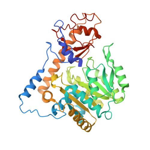
6DNA

Crystal structure of T110A mutant human Glutamate oxaloacetate transaminase 1 (GOT1)


Experimental Data Snapshot

  • Method: X-RAY DIFFRACTION
  • Resolution: 3.00 Å
  • R-Value Free: 0.211 
  • R-Value Work: 0.184 
  • R-Value Observed: 0.185 

Starting Model: experimental
View more details

wwPDB Validation   3D Report Full Report


Ligand Structure Quality Assessment 


This is version 1.3 of the entry. See complete history


Literature

Biochemical Characterization and Structure-Based Mutational Analysis Provide Insight into the Binding and Mechanism of Action of Novel Aspartate Aminotransferase Inhibitors.

Holt, M.C.Assar, Z.Beheshti Zavareh, R.Lin, L.Anglin, J.Mashadova, O.Haldar, D.Mullarky, E.Kremer, D.M.Cantley, L.C.Kimmelman, A.C.Stein, A.J.Lairson, L.L.Lyssiotis, C.A.

(2018) Biochemistry 57: 6604-6614

  • DOI: https://doi.org/10.1021/acs.biochem.8b00914
  • Primary Citation of Related Structures:  
    6DNA, 6DNB, 6DND

  • PubMed Abstract: 

    Pancreatic cancer cells are characterized by deregulated metabolic programs that facilitate growth and resistance to oxidative stress. Among these programs, pancreatic cancers preferentially utilize a metabolic pathway through the enzyme aspartate aminotransferase 1 [also known as glutamate oxaloacetate transaminase 1 (GOT1)] to support cellular redox homeostasis. As such, small molecule inhibitors that target GOT1 could serve as starting points for the development of new therapies for pancreatic cancer. We ran a high-throughput screen for inhibitors of GOT1 and identified a small molecule, iGOT1-01, with in vitro GOT1 inhibitor activity. Application in pancreatic cancer cells revealed metabolic and growth inhibitory activity reflecting a promiscuous inhibitory profile. We then performed an in silico docking analysis to study inhibitor-GOT1 interactions with iGOT1-01 analogues that possess improved solubility and potency properties. These results suggested that the GOT1 inhibitor competed for binding to the pyridoxal 5-phosphate (PLP) cofactor site of GOT1. To analyze how the GOT1 inhibitor bound to GOT1, a series of GOT1 mutant enzymes that abolished PLP binding were generated. Application of the mutants in X-ray crystallography and thermal shift assays again suggested but were unable to formally conclude that the GOT1 inhibitor bound to the PLP site. Mutational studies revealed the relationship between PLP binding and the thermal stability of GOT1 while highlighting the essential nature of several residues for GOT1 catalytic activity. Insight into the mode of action of GOT1 inhibitors may provide leads to the development of drugs that target redox balance in pancreatic cancer.


  • Organizational Affiliation

    Cayman Chemical Company , 1180 East Ellsworth , Ann Arbor , Michigan 48108 , United States.


Macromolecules
Find similar proteins by:  (by identity cutoff)  |  3D Structure
Entity ID: 1
MoleculeChains Sequence LengthOrganismDetailsImage
Aspartate aminotransferase, cytoplasmic
A, B, C, D, E
A, B, C, D, E, F
408Homo sapiensMutation(s): 1 
Gene Names: GOT1
EC: 2.6.1.1 (PDB Primary Data), 2.6.1.3 (PDB Primary Data)
UniProt & NIH Common Fund Data Resources
Find proteins for P17174 (Homo sapiens)
Explore P17174 
Go to UniProtKB:  P17174
PHAROS:  P17174
GTEx:  ENSG00000120053 
Entity Groups  
Sequence Clusters30% Identity50% Identity70% Identity90% Identity95% Identity100% Identity
UniProt GroupP17174
Sequence Annotations
Expand
  • Reference Sequence
Small Molecules
Ligands 2 Unique
IDChains Name / Formula / InChI Key2D Diagram3D Interactions
PLP
Query on PLP

Download Ideal Coordinates CCD File 
H [auth A]
J [auth B]
K [auth C]
M [auth D]
N [auth E]
H [auth A],
J [auth B],
K [auth C],
M [auth D],
N [auth E],
O [auth F]
PYRIDOXAL-5'-PHOSPHATE
C8 H10 N O6 P
NGVDGCNFYWLIFO-UHFFFAOYSA-N
PG4
Query on PG4

Download Ideal Coordinates CCD File 
G [auth A],
I [auth B],
L [auth D]
TETRAETHYLENE GLYCOL
C8 H18 O5
UWHCKJMYHZGTIT-UHFFFAOYSA-N
Experimental Data & Validation

Experimental Data

  • Method: X-RAY DIFFRACTION
  • Resolution: 3.00 Å
  • R-Value Free: 0.211 
  • R-Value Work: 0.184 
  • R-Value Observed: 0.185 
  • Space Group: C 1 2 1
Unit Cell:
Length ( Å )Angle ( ˚ )
a = 257.7α = 90
b = 148.626β = 89.98
c = 83.183γ = 90
Software Package:
Software NamePurpose
REFMACrefinement
HKL-2000data reduction
HKL-2000data scaling
MOLREPphasing

Structure Validation

View Full Validation Report


Ligand Structure Quality Assessment 


Entry History 

Deposition Data

Revision History  (Full details and data files)

  • Version 1.0: 2018-11-14
    Type: Initial release
  • Version 1.1: 2018-11-21
    Changes: Data collection, Database references
  • Version 1.2: 2018-12-05
    Changes: Data collection, Database references
  • Version 1.3: 2023-10-11
    Changes: Data collection, Database references, Refinement description