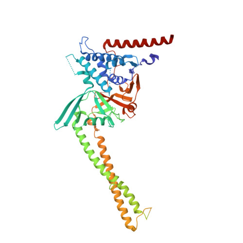
6HEJ

Structure of human USP28


Experimental Data Snapshot

  • Method: X-RAY DIFFRACTION
  • Resolution: 2.79 Å
  • R-Value Free: 0.286 
  • R-Value Work: 0.261 
  • R-Value Observed: 0.262 

Starting Model: experimental
View more details

wwPDB Validation   3D Report Full Report


This is version 1.3 of the entry. See complete history


Literature

Distinct USP25 and USP28 Oligomerization States Regulate Deubiquitinating Activity.

Gersch, M.Wagstaff, J.L.Toms, A.V.Graves, B.Freund, S.M.V.Komander, D.

(2019) Mol Cell 74: 436

  • DOI: https://doi.org/10.1016/j.molcel.2019.02.030
  • Primary Citation of Related Structures:  
    6HEH, 6HEI, 6HEJ, 6HEK, 6HEL, 6HEM

  • PubMed Abstract: 

    The evolutionarily related deubiquitinating enzymes (DUBs) USP25 and USP28 comprise an identical overall domain architecture but are functionally non-redundant: USP28 stabilizes c-MYC and other nuclear proteins, and USP25 regulates inflammatory TRAF signaling. We here compare molecular features of USP25 and USP28. Active enzymes form distinctively shaped dimers, with a dimerizing insertion spatially separating independently active catalytic domains. In USP25, but not USP28, two dimers can form an autoinhibited tetramer, where a USP25-specific, conserved insertion sequence blocks ubiquitin binding. In full-length enzymes, a C-terminal domain with a previously unknown fold has no impact on oligomerization, but N-terminal regions affect the dimer-tetramer equilibrium in vitro. We confirm oligomeric states of USP25 and USP28 in cells and show that modulating oligomerization affects substrate stabilization in accordance with in vitro activity data. Our work highlights how regions outside of the catalytic domain enable a conceptually intriguing interplay of DUB oligomerization and activity.


  • Organizational Affiliation

    Medical Research Council Laboratory of Molecular Biology, Francis Crick Avenue, Cambridge CB2 0QH, UK; Chemical Genomics Centre, Max-Planck-Institute of Molecular Physiology, Otto-Hahn-Str. 11, 44227 Dortmund, Germany; Department of Chemistry and Chemical Biology, Technical University Dortmund, Otto-Hahn-Str. 4a, 44227 Dortmund, Germany.


Macromolecules
Find similar proteins by:  (by identity cutoff)  |  3D Structure
Entity ID: 1
MoleculeChains Sequence LengthOrganismDetailsImage
Ubiquitin carboxyl-terminal hydrolase 28
A, B
556Homo sapiensMutation(s): 0 
Gene Names: USP28KIAA1515
EC: 3.4.19.12
UniProt & NIH Common Fund Data Resources
Find proteins for Q96RU2 (Homo sapiens)
Explore Q96RU2 
Go to UniProtKB:  Q96RU2
PHAROS:  Q96RU2
GTEx:  ENSG00000048028 
Entity Groups  
Sequence Clusters30% Identity50% Identity70% Identity90% Identity95% Identity100% Identity
UniProt GroupQ96RU2
Sequence Annotations
Expand
  • Reference Sequence
Experimental Data & Validation

Experimental Data

  • Method: X-RAY DIFFRACTION
  • Resolution: 2.79 Å
  • R-Value Free: 0.286 
  • R-Value Work: 0.261 
  • R-Value Observed: 0.262 
  • Space Group: I 2 2 2
Unit Cell:
Length ( Å )Angle ( ˚ )
a = 104.235α = 90
b = 200.455β = 90
c = 206.061γ = 90
Software Package:
Software NamePurpose
PHENIXrefinement
xia2data reduction
STARANISOdata scaling
PHASERphasing
autoPROCdata collection
Cootmodel building
RESTRAINrefinement

Structure Validation

View Full Validation Report


Entry History & Funding Information

Deposition Data


Funding OrganizationLocationGrant Number
Medical Research Council (United Kingdom)United KingdomU105192732
European Research CouncilUnited Kingdom724804

Revision History  (Full details and data files)

  • Version 1.0: 2019-03-27
    Type: Initial release
  • Version 1.1: 2019-04-10
    Changes: Data collection, Database references
  • Version 1.2: 2019-05-15
    Changes: Data collection, Database references
  • Version 1.3: 2024-01-17
    Changes: Data collection, Database references, Refinement description