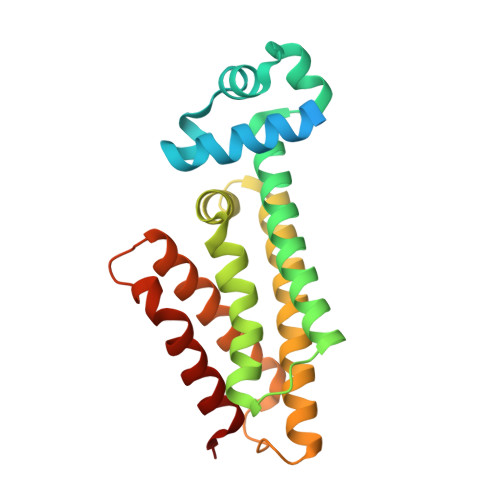
A comprehensive analysis of the protein-ligand interactions in crystal structures of Mycobacterium tuberculosis EthR.
Tanina, A., Wohlkonig, A., Soror, S.H., Flipo, M., Villemagne, B., Prevet, H., Deprez, B., Moune, M., Peree, H., Meyer, F., Baulard, A.R., Willand, N., Wintjens, R.(2018) Biochim Biophys Acta Proteins Proteom 1867: 248-258
- PubMed: 30553830
- DOI: https://doi.org/10.1016/j.bbapap.2018.12.003
- Primary Citation of Related Structures:
6HNX, 6HNZ, 6HO0, 6HO1, 6HO2, 6HO3, 6HO4, 6HO5, 6HO6, 6HO7, 6HO8, 6HO9, 6HOA, 6HOB, 6HOC, 6HOD, 6HOE, 6HOF - PubMed Abstract:
The Mycobacterium tuberculosis EthR is a member of the TetR family of repressors, controlling the expression of EthA, a mono-oxygenase responsible for the bioactivation of the prodrug ethionamide. This protein was established as a promising therapeutic target against tuberculosis, allowing, when inhibited by a drug-like molecule, to boost the action of ethionamide. Dozens of EthR crystal structures have been solved in complex with ligands. Herein, we disclose EthR structures in complex with 18 different small molecules and then performed in-depth analysis on the complete set of EthR structures that provides insights on EthR-ligand interactions. The 81 molecules solved in complex with EthR show a large diversity of chemical structures that were split up into several chemical clusters. Two of the most striking common points of EthR-ligand interactions are the quasi-omnipresence of a hydrogen bond bridging compounds with Asn 179 and the high occurrence of π-π interactions involving Phe 110 . A systematic analysis of the protein-ligand contacts identified eight hot spot residues that defined the basic structural features governing the binding mode of small molecules to EthR. Implications for the design of new potent inhibitors are discussed.
Organizational Affiliation:
Unité Microbiologie, Bioorganique et Macromoléculaire (CP206/04), département R3D, Faculté de Pharmacie, Université Libre de Bruxelles, B-1050 Brussels, Belgium.


