6J4Q

Structural basis of tubulin detyrosination by vasohibins-SVBP enzyme complex and functional implications


Experimental Data Snapshot

  • Method: X-RAY DIFFRACTION
  • Resolution: 2.70 Å
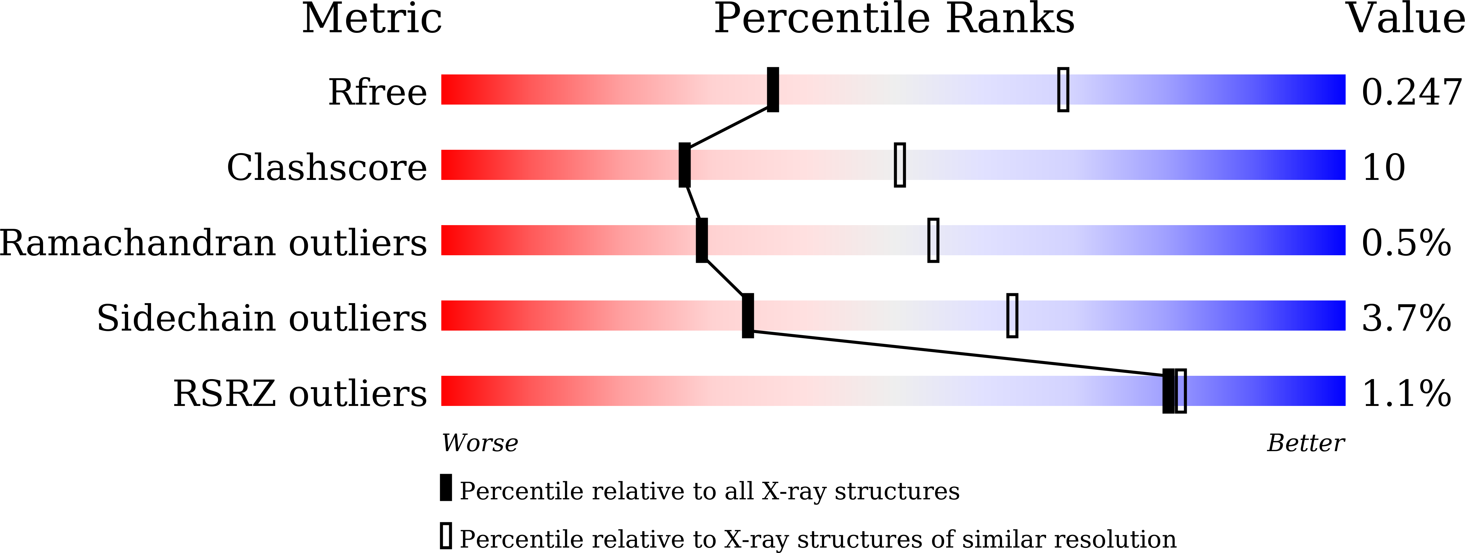
  • R-Value Free: 0.247 
  • R-Value Work: 0.183 
  • R-Value Observed: 0.187 

Starting Model: experimental
View more details

wwPDB Validation   3D Report Full Report


Ligand Structure Quality Assessment 


This is version 1.3 of the entry. See complete history


Literature

Structural basis of tubulin detyrosination by the vasohibin-SVBP enzyme complex.

Wang, N.Bosc, C.Ryul Choi, S.Boulan, B.Peris, L.Olieric, N.Bao, H.Krichen, F.Chen, L.Andrieux, A.Olieric, V.Moutin, M.J.Steinmetz, M.O.Huang, H.

(2019) Nat Struct Mol Biol 26: 571-582

  • DOI: https://doi.org/10.1038/s41594-019-0241-y
  • Primary Citation of Related Structures:  
    6J4O, 6J4P, 6J4Q, 6J4S, 6J4U, 6J4V, 6QBY

  • PubMed Abstract: 

    Vasohibins are tubulin tyrosine carboxypeptidases that are important in neuron physiology. We examined the crystal structures of human vasohibin 1 and 2 in complex with small vasohibin-binding protein (SVBP) in the absence and presence of different inhibitors and a C-terminal α-tubulin peptide. In combination with functional data, we propose that SVBP acts as an activator of vasohibins. An extended groove and a distinctive surface residue patch of vasohibins define the specific determinants for recognizing and cleaving the C-terminal tyrosine of α-tubulin and for binding microtubules, respectively. The vasohibin-SVBP interaction and the ability of the enzyme complex to associate with microtubules regulate axon specification of neurons. Our results define the structural basis of tubulin detyrosination by vasohibins and show the relevance of this process for neuronal development. Our findings offer a unique platform for developing drugs against human conditions with abnormal tubulin tyrosination levels, such as cancer, heart defects and possibly brain disorders.


  • Organizational Affiliation

    Department of Biology, Southern University of Science and Technology, Shenzhen, China.


Macromolecules
Find similar proteins by:  (by identity cutoff)  |  3D Structure
Entity ID: 1
MoleculeChains Sequence LengthOrganismDetailsImage
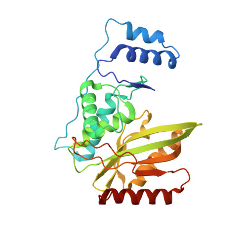
Tubulinyl-Tyr carboxypeptidase 2A,
C,
E [auth F],
G [auth J]
251Homo sapiensMutation(s): 0 
Gene Names: VASH2VASHL
EC: 3.4.17.17
UniProt & NIH Common Fund Data Resources
Find proteins for Q86V25 (Homo sapiens)
Explore Q86V25 
Go to UniProtKB:  Q86V25
PHAROS:  Q86V25
GTEx:  ENSG00000143494 
Entity Groups  
Sequence Clusters30% Identity50% Identity70% Identity90% Identity95% Identity100% Identity
UniProt GroupQ86V25
Sequence Annotations
Expand
  • Reference Sequence
Find similar proteins by:  (by identity cutoff)  |  3D Structure
Entity ID: 2
MoleculeChains Sequence LengthOrganismDetailsImage
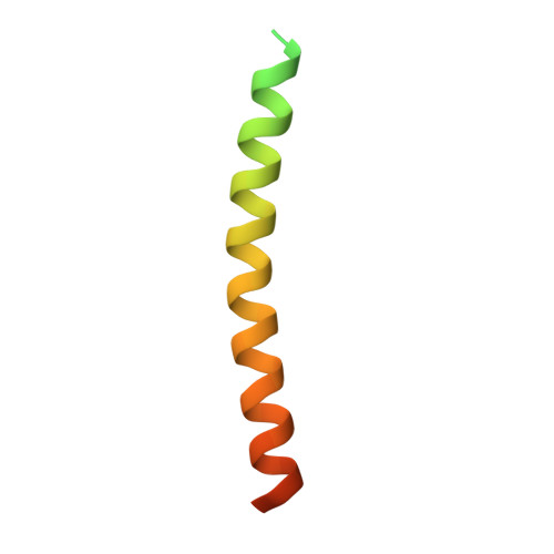
Small vasohibin-binding proteinB,
D,
F [auth G],
H [auth K]
66Homo sapiensMutation(s): 0 
Gene Names: SVBPCCDC23
UniProt & NIH Common Fund Data Resources
Find proteins for Q8N300 (Homo sapiens)
Explore Q8N300 
Go to UniProtKB:  Q8N300
PHAROS:  Q8N300
GTEx:  ENSG00000177868 
Entity Groups  
Sequence Clusters30% Identity50% Identity70% Identity90% Identity95% Identity100% Identity
UniProt GroupQ8N300
Sequence Annotations
Expand
  • Reference Sequence
Small Molecules
Ligands 2 Unique
IDChains Name / Formula / InChI Key2D Diagram3D Interactions
TQ8
Query on TQ8

Download Ideal Coordinates CCD File 
I [auth A]
L [auth B]
M [auth C]
O [auth F]
Q [auth G]
I [auth A],
L [auth B],
M [auth C],
O [auth F],
Q [auth G],
R [auth J]
N-[(2S)-4-chloro-3-oxo-1-phenyl-butan-2-yl]-4-methyl-benzenesulfonamide
C17 H18 Cl N O3 S
MQUQNUAYKLCRME-INIZCTEOSA-N
GOL
Query on GOL

Download Ideal Coordinates CCD File 
J [auth A],
K [auth A],
N [auth C],
P [auth F],
S [auth J]
GLYCEROL
C3 H8 O3
PEDCQBHIVMGVHV-UHFFFAOYSA-N
Experimental Data & Validation

Experimental Data

  • Method: X-RAY DIFFRACTION
  • Resolution: 2.70 Å
  • R-Value Free: 0.247 
  • R-Value Work: 0.183 
  • R-Value Observed: 0.187 
  • Space Group: P 32 2 1
Unit Cell:
Length ( Å )Angle ( ˚ )
a = 117.177α = 90
b = 117.177β = 90
c = 209.044γ = 120
Software Package:
Software NamePurpose
PHENIXrefinement
HKL-3000data reduction
HKL-3000data scaling
PHASERphasing

Structure Validation

View Full Validation Report


Ligand Structure Quality Assessment 


Entry History & Funding Information

Deposition Data


Funding OrganizationLocationGrant Number
Ministry of Science and Technology (China)China2018YFC1004500
National Natural Science Foundation of ChinaChinathe Thousand Young Talents Program
National Natural Science Foundation of ChinaChinaK18221002

Revision History  (Full details and data files)

  • Version 1.0: 2019-05-01
    Type: Initial release
  • Version 1.1: 2019-11-13
    Changes: Database references
  • Version 1.2: 2023-11-22
    Changes: Data collection, Database references, Refinement description
  • Version 1.3: 2024-10-23
    Changes: Structure summary