6MB7

Binary (paromomycin) structure of AAC-IIIb


Experimental Data Snapshot

  • Method: X-RAY DIFFRACTION
  • Resolution: 2.20 Å
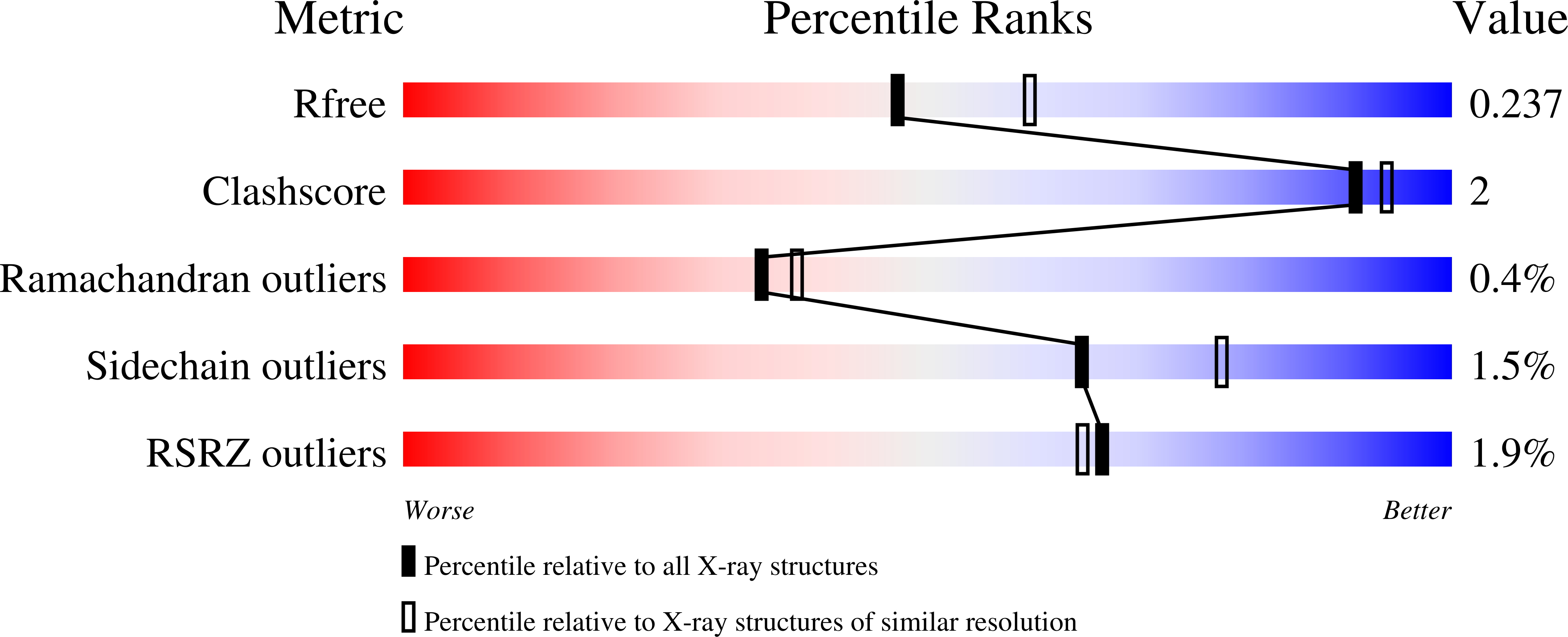
  • R-Value Free: 0.237 
  • R-Value Work: 0.200 
  • R-Value Observed: 0.202 

Starting Model: experimental
View more details

wwPDB Validation   3D Report Full Report


Ligand Structure Quality Assessment 


This is version 1.2 of the entry. See complete history


Literature

Encoding of Promiscuity in an Aminoglycoside Acetyltransferase.

Kumar, P.Selvaraj, B.Serpersu, E.H.Cuneo, M.J.

(2018) J Med Chem 61: 10218-10227

  • DOI: https://doi.org/10.1021/acs.jmedchem.8b01393
  • Primary Citation of Related Structures:  
    6MB4, 6MB5, 6MB6, 6MB7, 6MB8, 6MB9

  • PubMed Abstract: 

    Aminoglycoside antibiotics are a large family of antibiotics that can be divided into two distinct classes on the basis of the substitution pattern of the central deoxystreptamine ring. Although aminoglycosides are chemically, structurally, and topologically diverse, some aminoglycoside-modifying enzymes (AGMEs) are able to inactivate as many as 15 aminoglycosides from the two main classes, the kanamycin- and neomycin-based antibiotics. Here, we present the crystal structure of a promiscuous AGME, aminoglycoside- N3-acetyltransferase-IIIb (AAC-IIIb), in the apo form, in binary drug (sisomicin, neomycin, and paromomycin) and coenzyme A (CoASH) complexes, and in the ternary neomycin-CoASH complex. These data provide a structural framework for interpretation of the thermodynamics of enzyme-ligand interactions and the role of solvent in the recognition of ligands. In combination with the recent structure of an AGME that does not have broad substrate specificity, these structures allow for the direct determination of how antibiotic promiscuity is encoded in some AGMEs.


  • Organizational Affiliation

    Graduate School of Genome Science and Technology , The University of Tennessee and Oak Ridge National Laboratory , 1414 West Cumberland Avenue , Knoxville , Tennessee 37996 , United States.


Macromolecules
Find similar proteins by:  (by identity cutoff)  |  3D Structure
Entity ID: 1
MoleculeChains Sequence LengthOrganismDetailsImage
Aac(3)-IIIb protein274Pseudomonas aeruginosaMutation(s): 0 
Gene Names: aac(3)-IIIb
EC: 2.3.1
UniProt
Find proteins for Q51405 (Pseudomonas aeruginosa)
Explore Q51405 
Go to UniProtKB:  Q51405
Entity Groups  
Sequence Clusters30% Identity50% Identity70% Identity90% Identity95% Identity100% Identity
UniProt GroupQ51405
Sequence Annotations
Expand
  • Reference Sequence
Small Molecules
Ligands 1 Unique
IDChains Name / Formula / InChI Key2D Diagram3D Interactions
PAR
Query on PAR

Download Ideal Coordinates CCD File 
B [auth A]PAROMOMYCIN
C23 H45 N5 O14
UOZODPSAJZTQNH-LSWIJEOBSA-N
Experimental Data & Validation

Experimental Data

  • Method: X-RAY DIFFRACTION
  • Resolution: 2.20 Å
  • R-Value Free: 0.237 
  • R-Value Work: 0.200 
  • R-Value Observed: 0.202 
  • Space Group: P 32 2 1
Unit Cell:
Length ( Å )Angle ( ˚ )
a = 106.19α = 90
b = 106.19β = 90
c = 69γ = 120
Software Package:
Software NamePurpose
PHENIXrefinement
Aimlessdata scaling
PDB_EXTRACTdata extraction
MOSFLMdata reduction
PHASERphasing

Structure Validation

View Full Validation Report


Ligand Structure Quality Assessment 


Entry History 

Deposition Data

Revision History  (Full details and data files)

  • Version 1.0: 2018-11-07
    Type: Initial release
  • Version 1.1: 2018-12-05
    Changes: Data collection, Database references
  • Version 1.2: 2023-10-11
    Changes: Data collection, Database references, Refinement description, Structure summary