6N5C | pdb_00006n5c

Crystal structure of the catalytic domain of PPIP5K2 in complex with AMPPNP and 5-PCF2Am-InsP5


Experimental Data Snapshot

  • Method: X-RAY DIFFRACTION
  • Resolution: 1.95 Å
  • R-Value Free: 
    0.214 (Depositor), 0.220 (DCC) 
  • R-Value Work: 
    0.175 (Depositor), 0.180 (DCC) 
  • R-Value Observed: 
    0.178 (Depositor) 

Starting Model: experimental
View more details

wwPDB Validation   3D Report Full Report


Ligand Structure Quality Assessment 

Created with Raphaël 2.3.0Worse 01 BetterLigand structure goodness of fit to experimental dataBest fitted KDJClick on this verticalbar to view detailsBest fitted ANPClick on this verticalbar to view details

This is version 1.2 of the entry. See complete history


Literature

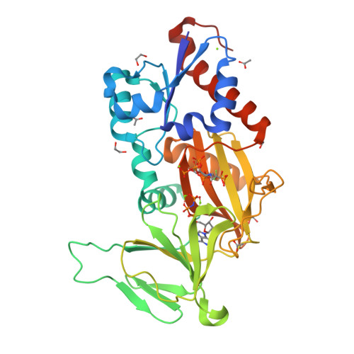
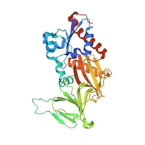
Synthesis of an alpha-phosphono-alpha , alpha-difluoroacetamide analogue of the diphosphoinositol pentakisphosphate 5-InsP7.

Riley, A.M.Wang, H.Shears, S.B.Potter, B.V.L.

(2019) Medchemcomm 10: 1165-1172

  • DOI: https://doi.org/10.1039/c9md00163h
  • Primary Citation of Related Structures:  
    6N5C

  • PubMed Abstract: 

    Diphosphoinositol phosphates (PP-InsPs) are an evolutionarily ancient group of signalling molecules that are essential to cellular and organismal homeostasis. As the detailed mechanisms of PP-InsP signalling begin to emerge, synthetic analogues of PP-InsPs containing stabilised mimics of the labile diphosphate group can provide valuable investigational tools. We synthesised 5-PCF 2 Am-InsP 5 ( 1 ), a novel fluorinated phosphonate analogue of 5-PP-InsP 5 , and obtained an X-ray crystal structure of 1 in complex with diphosphoinositol pentakisphosphate kinase 2 (PPIP5K2). 5-PCF 2 Am-InsP 5 binds to the kinase domain of PPIP5K2 in a similar orientation to that of the natural substrate 5-PP-InsP 5 and the PCF 2 Am structure can mimic many aspects of the diphosphate group in 5-PP-InsP 5 . We propose that 1 , the structural and electronic properties of which are in some ways complementary to those of existing phosphonoacetate and methylenebisphosphonate analogues of 5-PP-InsP 5 , may be a useful addition to the expanding array of chemical tools for the investigation of signalling by PP-InsPs. The PCF 2 Am group may also deserve attention for wider application as a diphosphate mimic.


  • Organizational Affiliation

    Medicinal Chemistry and Drug Discovery , Department of Pharmacology , University of Oxford , Mansfield Road , Oxford OX1 3QT , UK . Email: barry.potter@pharm.ox.ac.uk ; ; Tel: +44 (0)1865 271945.


Macromolecules
Find similar proteins by:  (by identity cutoff)  |  3D Structure
Entity ID: 1
MoleculeChains Sequence LengthOrganismDetailsImage
Inositol hexakisphosphate and diphosphoinositol-pentakisphosphate kinase 2330Homo sapiensMutation(s): 0 
Gene Names: PPIP5K2HISPPD1KIAA0433VIP2
EC: 2.7.4.21 (PDB Primary Data), 2.7.4.24 (PDB Primary Data)
UniProt & NIH Common Fund Data Resources
Find proteins for O43314 (Homo sapiens)
Explore O43314 
Go to UniProtKB:  O43314
PHAROS:  O43314
GTEx:  ENSG00000145725 
Entity Groups  
Sequence Clusters30% Identity50% Identity70% Identity90% Identity95% Identity100% Identity
UniProt GroupO43314
Sequence Annotations
Expand
  • Reference Sequence
Small Molecules
Ligands 5 Unique
IDChains Name / Formula / InChI Key2D Diagram3D Interactions
KDJ
Query on KDJ

Download Ideal Coordinates CCD File 
G [auth A](1,1-difluoro-2-oxo-2-{[(1s,2R,3S,4s,5R,6S)-2,3,4,5,6-pentakis(phosphonooxy)cyclohexyl]amino}ethyl)phosphonic acid
C8 H19 F2 N O24 P6
XBHZOGSBYRIXJA-QWBQGLJISA-N
ANP
Query on ANP

Download Ideal Coordinates CCD File 
B [auth A]PHOSPHOAMINOPHOSPHONIC ACID-ADENYLATE ESTER
C10 H17 N6 O12 P3
PVKSNHVPLWYQGJ-KQYNXXCUSA-N
EDO
Query on EDO

Download Ideal Coordinates CCD File 
I [auth A],
K [auth A],
L [auth A],
M [auth A],
O [auth A]
1,2-ETHANEDIOL
C2 H6 O2
LYCAIKOWRPUZTN-UHFFFAOYSA-N
ACT
Query on ACT

Download Ideal Coordinates CCD File 
H [auth A],
J [auth A],
N [auth A]
ACETATE ION
C2 H3 O2
QTBSBXVTEAMEQO-UHFFFAOYSA-M
MG
Query on MG

Download Ideal Coordinates CCD File 
C [auth A],
D [auth A],
E [auth A],
F [auth A]
MAGNESIUM ION
Mg
JLVVSXFLKOJNIY-UHFFFAOYSA-N
Experimental Data & Validation

Experimental Data

  • Method: X-RAY DIFFRACTION
  • Resolution: 1.95 Å
  • R-Value Free:  0.214 (Depositor), 0.220 (DCC) 
  • R-Value Work:  0.175 (Depositor), 0.180 (DCC) 
  • R-Value Observed: 0.178 (Depositor) 
Space Group: P 21 21 21
Unit Cell:
Length ( Å )Angle ( ˚ )
a = 88.604α = 90
b = 111.032β = 90
c = 41.429γ = 90
Software Package:
Software NamePurpose
REFMACrefinement
HKL-2000data reduction
HKL-2000data scaling
REFMACphasing

Structure Validation

View Full Validation Report


Ligand Structure Quality Assessment 

Created with Raphaël 2.3.0Worse 01 BetterLigand structure goodness of fit to experimental dataBest fitted KDJClick on this verticalbar to view detailsBest fitted ANPClick on this verticalbar to view details

Entry History & Funding Information

Deposition Data


Funding OrganizationLocationGrant Number
National Institutes of Health/National Institute of Environmental Health Sciences (NIH/NIEHS)United States1ZIAES080046-29

Revision History  (Full details and data files)

  • Version 1.0: 2019-08-21
    Type: Initial release
  • Version 1.1: 2019-12-18
    Changes: Author supporting evidence
  • Version 1.2: 2023-10-11
    Changes: Data collection, Database references, Derived calculations, Refinement description