6P11

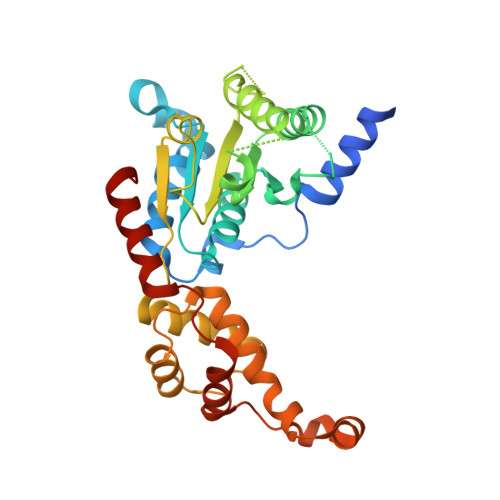
Structure of spastin AAA domain (T692A mutant) in complex with JNJ-7706621 inhibitor


Experimental Data Snapshot

  • Method: X-RAY DIFFRACTION
  • Resolution: 2.15 Å
  • R-Value Free: 0.241 
  • R-Value Work: 0.213 
  • R-Value Observed: 0.215 

Starting Model: experimental
View more details

wwPDB Validation   3D Report Full Report

Currently 6P11 does not have a validation slider image.


This is version 1.3 of the entry. See complete history


Literature

Analyzing Resistance to Design Selective Chemical Inhibitors for AAA Proteins.

Pisa, R.Cupido, T.Steinman, J.B.Jones, N.H.Kapoor, T.M.

(2019) Cell Chem Biol 26: 1263

  • DOI: https://doi.org/10.1016/j.chembiol.2019.06.001
  • Primary Citation of Related Structures:  
    6P10, 6P11, 6P12, 6P13, 6P14

  • PubMed Abstract: 

    Drug-like inhibitors are often designed by mimicking cofactor or substrate interactions with enzymes. However, as active sites are comprised of conserved residues, it is difficult to identify the critical interactions needed to design selective inhibitors. We are developing an approach, named RADD (resistance analysis during design), which involves engineering point mutations in the target to generate active alleles and testing compounds against them. Mutations that alter compound potency identify residues that make key interactions with the inhibitor and predict target-binding poses. Here, we apply this approach to analyze how diaminotriazole-based inhibitors bind spastin, a microtubule-severing AAA (ATPase associated with diverse cellular activities) protein. The distinct binding poses predicted for two similar inhibitors were confirmed by a series of X-ray structures. Importantly, our approach not only reveals how selective inhibition of the target can be achieved but also identifies resistance-conferring mutations at the early stages of the design process.


  • Organizational Affiliation

    Laboratory of Chemistry and Cell Biology, The Rockefeller University, New York, NY 10065, USA; Tri-Institutional PhD Program in Chemical Biology, The Rockefeller University, New York, NY 10065, USA.


Macromolecules
Find similar proteins by:  (by identity cutoff)  |  3D Structure
Entity ID: 1
MoleculeChains Sequence LengthOrganismDetailsImage
Drosophila melanogaster Spastin AAA domainA [auth B]314Drosophila melanogasterMutation(s): 1 
Gene Names: spasCG5977
EC: 5.6.1.1
UniProt
Find proteins for Q8I0P1 (Drosophila melanogaster)
Explore Q8I0P1 
Go to UniProtKB:  Q8I0P1
Entity Groups  
Sequence Clusters30% Identity50% Identity70% Identity90% Identity95% Identity100% Identity
UniProt GroupQ8I0P1
Sequence Annotations
Expand
  • Reference Sequence
Small Molecules
Ligands 3 Unique
IDChains Name / Formula / InChI Key2D Diagram3D Interactions
SKE (Subject of Investigation/LOI)
Query on SKE

Download Ideal Coordinates CCD File 
C [auth B]4-({5-amino-1-[(2,6-difluorophenyl)carbonyl]-1H-1,2,4-triazol-3-yl}amino)benzenesulfonamide
C15 H12 F2 N6 O3 S
KDKUVYLMPJIGKA-UHFFFAOYSA-N
MPD
Query on MPD

Download Ideal Coordinates CCD File 
D [auth B](4S)-2-METHYL-2,4-PENTANEDIOL
C6 H14 O2
SVTBMSDMJJWYQN-YFKPBYRVSA-N
SO4
Query on SO4

Download Ideal Coordinates CCD File 
B
SULFATE ION
O4 S
QAOWNCQODCNURD-UHFFFAOYSA-L
Experimental Data & Validation

Experimental Data

  • Method: X-RAY DIFFRACTION
  • Resolution: 2.15 Å
  • R-Value Free: 0.241 
  • R-Value Work: 0.213 
  • R-Value Observed: 0.215 
  • Space Group: P 65
Unit Cell:
Length ( Å )Angle ( ˚ )
a = 79.446α = 90
b = 79.446β = 90
c = 97.171γ = 120
Software Package:
Software NamePurpose
HKL-2000data reduction
SCALEPACKdata scaling
PHENIXrefinement
PDB_EXTRACTdata extraction
PHASERphasing

Structure Validation

View Full Validation Report

Currently 6P11 does not have a validation slider image.


Entry History & Funding Information

Deposition Data


Funding OrganizationLocationGrant Number
National Institutes of Health/National Institute of General Medical Sciences (NIH/NIGMS)United StatesGM98579
National Institutes of Health/National Institute of General Medical Sciences (NIH/NIGMS)United StatesR35 GM130234-01

Revision History  (Full details and data files)

  • Version 1.0: 2019-07-24
    Type: Initial release
  • Version 1.1: 2019-10-02
    Changes: Data collection, Database references
  • Version 1.2: 2020-01-01
    Changes: Author supporting evidence
  • Version 1.3: 2023-10-11
    Changes: Data collection, Database references, Refinement description