6P1X

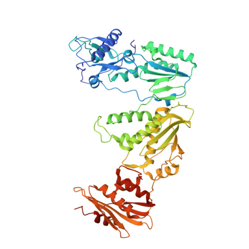
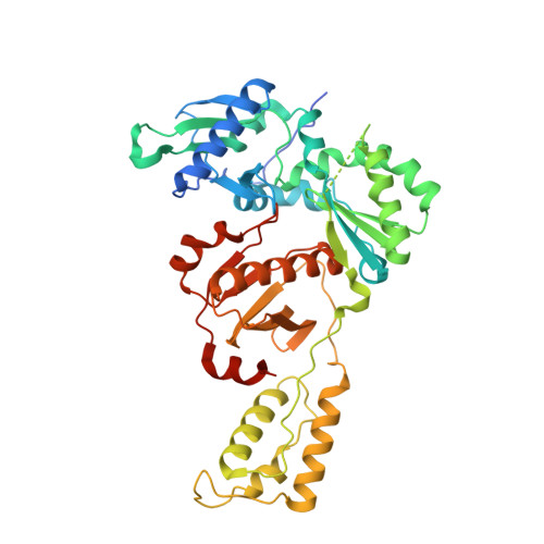
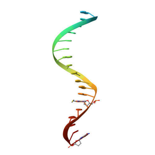
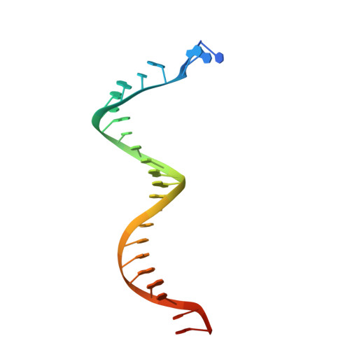
Structure of HIV-1 Reverse Transcriptase (RT) in complex with dsDNA and L-ddCTP


Experimental Data Snapshot

  • Method: X-RAY DIFFRACTION
  • Resolution: 2.55 Å
  • R-Value Free: 0.230 
  • R-Value Work: 0.188 
  • R-Value Observed: 0.190 

Starting Model: experimental
View more details

wwPDB Validation   3D Report Full Report


Ligand Structure Quality Assessment 


This is version 1.5 of the entry. See complete history


Literature

Structural insights into the recognition of nucleoside reverse transcriptase inhibitors by HIV-1 reverse transcriptase: First crystal structures with reverse transcriptase and the active triphosphate forms of lamivudine and emtricitabine.

Bertoletti, N.Chan, A.H.Schinazi, R.F.Yin, Y.W.Anderson, K.S.

(2019) Protein Sci 28: 1664-1675

  • DOI: https://doi.org/10.1002/pro.3681
  • Primary Citation of Related Structures:  
    6OR7, 6OTZ, 6OUN, 6P1I, 6P1X, 6P2G

  • PubMed Abstract: 

    The retrovirus HIV-1 has been a major health issue since its discovery in the early 80s. In 2017, over 37 million people were infected with HIV-1, of which 1.8 million were new infections that year. Currently, the most successful treatment regimen is the highly active antiretroviral therapy (HAART), which consists of a combination of three to four of the current 26 FDA-approved HIV-1 drugs. Half of these drugs target the reverse transcriptase (RT) enzyme that is essential for viral replication. One class of RT inhibitors is nucleoside reverse transcriptase inhibitors (NRTIs), a crucial component of the HAART. Once incorporated into DNA, NRTIs function as a chain terminator to stop viral DNA replication. Unfortunately, treatment with NRTIs is sometimes linked to toxicity caused by off-target side effects. NRTIs may also target the replicative human mitochondrial DNA polymerase (Pol γ), causing long-term severe drug toxicity. The goal of this work is to understand the discrimination mechanism of different NRTI analogues by RT. Crystal structures and kinetic experiments are essential for the rational design of new molecules that are able to bind selectively to RT and not Pol γ. Structural comparison of NRTI-binding modes with both RT and Pol γ enzymes highlights key amino acids that are responsible for the difference in affinity of these drugs to their targets. Therefore, the long-term goal of this research is to develop safer, next generation therapeutics that can overcome off-target toxicity.


  • Organizational Affiliation

    Department of Pharmacology, Yale University School of Medicine, New Haven, Connecticut.


Macromolecules

Find similar proteins by:  (by identity cutoff)  |  3D Structure
Entity ID: 1
MoleculeChains Sequence LengthOrganismDetailsImage
Reverse transcriptase/ribonuclease H560HIV-1 M:B_HXB2RMutation(s): 2 
Gene Names: gag-pol
EC: 2.7.7.49 (PDB Primary Data), 2.7.7.7 (PDB Primary Data), 3.1.26.13 (PDB Primary Data)
UniProt
Find proteins for P04585 (Human immunodeficiency virus type 1 group M subtype B (isolate HXB2))
Explore P04585 
Go to UniProtKB:  P04585
Entity Groups  
Sequence Clusters30% Identity50% Identity70% Identity90% Identity95% Identity100% Identity
UniProt GroupP04585
Sequence Annotations
Expand
  • Reference Sequence
Find similar proteins by:  (by identity cutoff)  |  3D Structure
Entity ID: 2
MoleculeChains Sequence LengthOrganismDetailsImage
p51 RT452HIV-1 M:B_HXB2RMutation(s): 1 
Gene Names: gag-pol
UniProt
Find proteins for P04585 (Human immunodeficiency virus type 1 group M subtype B (isolate HXB2))
Explore P04585 
Go to UniProtKB:  P04585
Entity Groups  
Sequence Clusters30% Identity50% Identity70% Identity90% Identity95% Identity100% Identity
UniProt GroupP04585
Sequence Annotations
Expand
  • Reference Sequence

Find similar nucleic acids by:  Sequence   |   3D Structure  

Entity ID: 3
MoleculeChains LengthOrganismImage
DNA Primer 20-merC [auth P]21synthetic construct
Sequence Annotations
Expand
  • Reference Sequence
Find similar nucleic acids by:  (by identity cutoff)  |  3D Structure
Entity ID: 4
MoleculeChains LengthOrganismImage
DNA template 27-merD [auth T]27synthetic construct
Sequence Annotations
Expand
  • Reference Sequence
Experimental Data & Validation

Experimental Data

  • Method: X-RAY DIFFRACTION
  • Resolution: 2.55 Å
  • R-Value Free: 0.230 
  • R-Value Work: 0.188 
  • R-Value Observed: 0.190 
  • Space Group: C 2 2 21
Unit Cell:
Length ( Å )Angle ( ˚ )
a = 169.213α = 90
b = 171.502β = 90
c = 107.306γ = 90
Software Package:
Software NamePurpose
PHENIXrefinement
XDSdata reduction
XDSdata scaling
PHASERphasing

Structure Validation

View Full Validation Report


Ligand Structure Quality Assessment 


Entry History & Funding Information

Deposition Data


Funding OrganizationLocationGrant Number
National Institutes of Health/National Institute of General Medical Sciences (NIH/NIGMS)United StatesGM049551

Revision History  (Full details and data files)

  • Version 1.0: 2019-07-24
    Type: Initial release
  • Version 1.1: 2019-08-21
    Changes: Data collection, Database references
  • Version 1.2: 2019-08-28
    Changes: Data collection, Database references
  • Version 1.3: 2020-01-01
    Changes: Author supporting evidence
  • Version 1.4: 2023-10-11
    Changes: Data collection, Database references, Derived calculations, Refinement description
  • Version 1.5: 2024-10-09
    Changes: Structure summary