6PA6

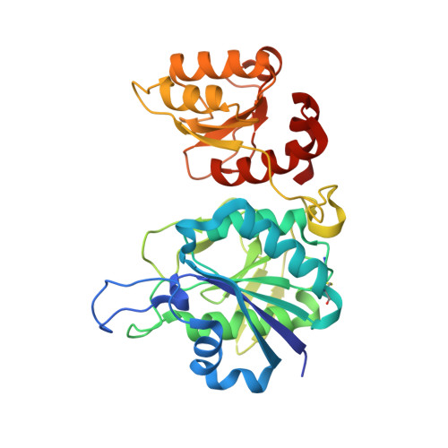
ECAII(T89V,K162T) MUTANT IN COMPLEX WITH L-ASN AT PH 8.3 in space group C2


Experimental Data Snapshot

  • Method: X-RAY DIFFRACTION
  • Resolution: 2.12 Å
  • R-Value Free: 0.231 
  • R-Value Work: 0.163 
  • R-Value Observed: 0.165 

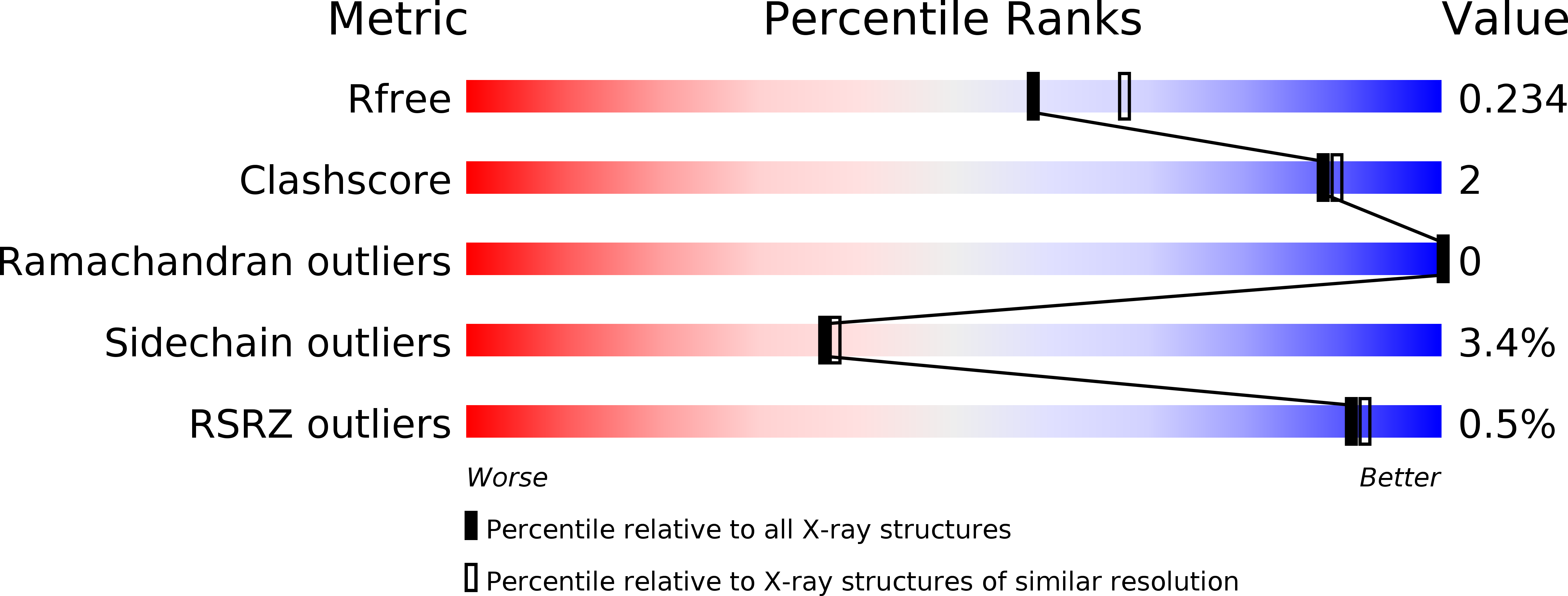
wwPDB Validation   3D Report Full Report


Ligand Structure Quality Assessment 


This is version 1.3 of the entry. See complete history


Literature

Geometric considerations support the double-displacement catalytic mechanism of l-asparaginase.

Lubkowski, J.Wlodawer, A.

(2019) Protein Sci 28: 1850-1864

  • DOI: https://doi.org/10.1002/pro.3709
  • Primary Citation of Related Structures:  
    6PA2, 6PA3, 6PA4, 6PA5, 6PA6, 6PA8, 6PA9, 6PAA, 6PAB, 6PAC, 6PAE

  • PubMed Abstract: 

    Twenty crystal structures of the complexes of l-asparaginase with l-Asn, l-Asp, and succinic acid that are currently available in the Protein Data Bank, as well as 11 additional structures determined in the course of this project, were analyzed in order to establish the level of conservation of the geometric parameters describing interactions between the substrates and the active site of the enzymes. We found that such stereochemical relationships are highly conserved, regardless of the organism from which the enzyme was isolated, specific crystallization conditions, or the nature of the ligands. Analysis of the geometry of the interactions, including Bürgi-Dunitz and Flippin-Lodge angles, indicated that Thr12 (Escherichia coli asparaginase II numbering) is optimally placed to be the primary nucleophile in the most likely scenario utilizing a double-displacement mechanism, whereas catalysis through a single-displacement mechanism appears to be the least likely.


  • Organizational Affiliation

    Macromolecular Crystallography Laboratory, National Cancer Institute, Frederick, Maryland.


Macromolecules
Find similar proteins by:  (by identity cutoff)  |  3D Structure
Entity ID: 1
MoleculeChains Sequence LengthOrganismDetailsImage
L-asparaginase 2
A, B, C, D
334Escherichia coli K-12Mutation(s): 2 
Gene Names: ansBb2957JW2924
EC: 3.5.1.1
UniProt
Find proteins for P00805 (Escherichia coli (strain K12))
Go to UniProtKB:  P00805
Sequence Annotations
Expand
  • Reference Sequence
Experimental Data & Validation

Experimental Data

  • Method: X-RAY DIFFRACTION
  • Resolution: 2.12 Å
  • R-Value Free: 0.231 
  • R-Value Work: 0.163 
  • R-Value Observed: 0.165 
  • Space Group: C 1 2 1
Unit Cell:
Length ( Å )Angle ( ˚ )
a = 151.22α = 90
b = 62.397β = 117.71
c = 140.875γ = 90
Software Package:
Software NamePurpose
HKL-2000data reduction
HKL-2000data scaling
PHASERphasing
REFMACrefinement
PDB_EXTRACTdata extraction

Structure Validation

View Full Validation Report


Ligand Structure Quality Assessment 


Entry History 

Deposition Data

Revision History  (Full details and data files)

  • Version 1.0: 2019-09-04
    Type: Initial release
  • Version 1.1: 2019-09-25
    Changes: Data collection, Database references
  • Version 1.2: 2019-10-02
    Changes: Data collection, Database references
  • Version 1.3: 2024-10-30
    Changes: Data collection, Database references, Structure summary