6PQI

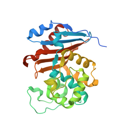
Crystal Structure of Class D Beta-lactamase OXA-48 with Cefotaxime


Experimental Data Snapshot

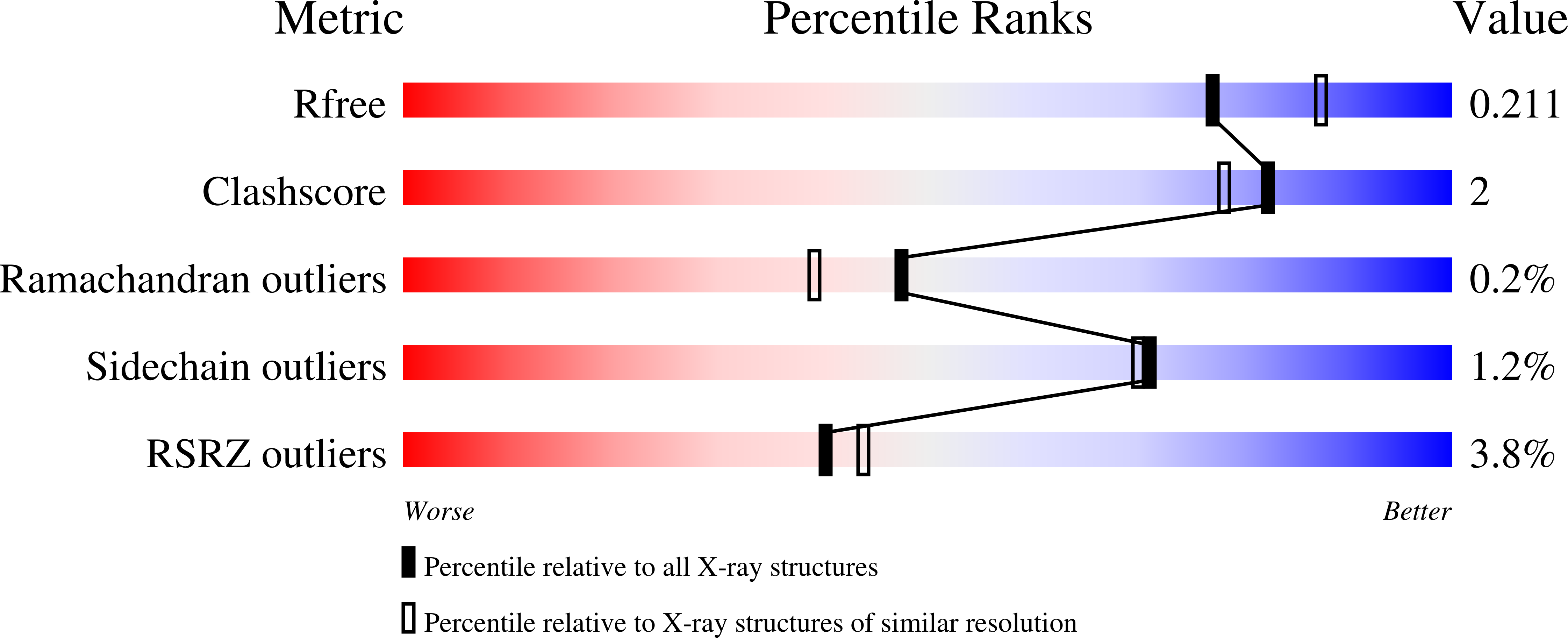
  • Method: X-RAY DIFFRACTION
  • Resolution: 2.05 Å
  • R-Value Free: 0.212 
  • R-Value Work: 0.182 
  • R-Value Observed: 0.183 

Starting Model: experimental
View more details

wwPDB Validation   3D Report Full Report


Ligand Structure Quality Assessment 


This is version 1.3 of the entry. See complete history


Literature

Structural Basis for Substrate Specificity and Carbapenemase Activity of OXA-48 Class D beta-Lactamase.

Akhtar, A.Pemberton, O.A.Chen, Y.

(2020) ACS Infect Dis 6: 261-271

  • DOI: https://doi.org/10.1021/acsinfecdis.9b00304
  • Primary Citation of Related Structures:  
    6PK0, 6PQI, 6PSG, 6PT1, 6PT5, 6PTU

  • PubMed Abstract: 

    Carbapenem-hydrolyzing class D β-lactamases (CHDLs) are a diverse family of enzymes that are rapidly becoming the predominant cause of bacterial resistance against β-lactam antibiotics in many regions of the world. OXA-48, an atypical member of CHDLs, is one of the most frequently observed in the clinic and exhibits a unique substrate profile. We applied X-ray crystallography to OXA-48 complexes with multiple β-lactam antibiotics to elucidate this enzyme's carbapenemase activity and its preference of imipenem over meropenem and other substrates such as cefotaxime. In particular, we obtained acyl-enzyme complexes of OXA-48 with imipenem, meropenem, faropenem, cefotaxime, and cefoxitin, and a product complex with imipenem. Importantly, the product complex captures a key reaction milestone with the newly generated carboxylate group still in the oxyanion hole, and represents the first such complex with a wild-type serine β-lactamase. A potential hydrogen bond is observed between the two carboxylate groups from the product and the carbamylated Lys73, representing the stage immediately after the breakage of the acyl-enzyme bond where the product carboxylate would be neutral. The placement of the product carboxylate also illustrates the approximate transient location of the deacylation water that has long eluded structural characterization in class D β-lactamases. Additionally, comparing the product complex with the acyl-enzyme intermediates provides new insights into the various mechanisms by which specific side chain groups hinder the access of the deacylation water to the acyl-enzyme linkage, especially in meropenem. Taken together, these data offer valuable information on the substrate specificity of OXA-48 and the catalytic mechanism of CHDLs.


  • Organizational Affiliation

    Department of Molecular Medicine , University of South Florida Morsani College of Medicine , 12901 Bruce B. Downs Boulevard, MDC 3522 , Tampa , Florida 33612 , United States.


Macromolecules
Find similar proteins by:  (by identity cutoff)  |  3D Structure
Entity ID: 1
MoleculeChains Sequence LengthOrganismDetailsImage
Class D Carbapenemase OXA-48
A, B, C, D
263Klebsiella pneumoniaeMutation(s): 0 
Gene Names: blaOXA
EC: 3.5.2.6
UniProt
Find proteins for Q6XEC0 (Klebsiella pneumoniae)
Explore Q6XEC0 
Go to UniProtKB:  Q6XEC0
Entity Groups  
Sequence Clusters30% Identity50% Identity70% Identity90% Identity95% Identity100% Identity
UniProt GroupQ6XEC0
Sequence Annotations
Expand
  • Reference Sequence
Small Molecules
Ligands 3 Unique
IDChains Name / Formula / InChI Key2D Diagram3D Interactions
CEF (Subject of Investigation/LOI)
Query on CEF

Download Ideal Coordinates CCD File 
E [auth A],
I [auth B],
K [auth C],
N [auth D]
CEFOTAXIME, C3' cleaved, open, bound form
C14 H15 N5 O5 S2
NRYMPLKBKFIWQC-YVCCLBOHSA-N
ACT
Query on ACT

Download Ideal Coordinates CCD File 
H [auth A],
M [auth C],
Q [auth D]
ACETATE ION
C2 H3 O2
QTBSBXVTEAMEQO-UHFFFAOYSA-M
CL
Query on CL

Download Ideal Coordinates CCD File 
F [auth A]
G [auth A]
J [auth B]
L [auth C]
O [auth D]
F [auth A],
G [auth A],
J [auth B],
L [auth C],
O [auth D],
P [auth D]
CHLORIDE ION
Cl
VEXZGXHMUGYJMC-UHFFFAOYSA-M
Experimental Data & Validation

Experimental Data

  • Method: X-RAY DIFFRACTION
  • Resolution: 2.05 Å
  • R-Value Free: 0.212 
  • R-Value Work: 0.182 
  • R-Value Observed: 0.183 
  • Space Group: P 1 21 1
Unit Cell:
Length ( Å )Angle ( ˚ )
a = 59.732α = 90
b = 107.005β = 104.17
c = 96.196γ = 90
Software Package:
Software NamePurpose
PHENIXrefinement
HKL-2000data scaling
PHASERphasing
PDB_EXTRACTdata extraction
HKL-2000data reduction

Structure Validation

View Full Validation Report


Ligand Structure Quality Assessment 


Entry History & Funding Information

Deposition Data


Funding OrganizationLocationGrant Number
National Institutes of Health/National Institute Of Allergy and Infectious Diseases (NIH/NIAID)United StatesAI103158

Revision History  (Full details and data files)

  • Version 1.0: 2020-01-22
    Type: Initial release
  • Version 1.1: 2020-08-05
    Changes: Database references
  • Version 1.2: 2023-10-11
    Changes: Data collection, Database references, Refinement description
  • Version 1.3: 2024-10-23
    Changes: Structure summary