6TRQ

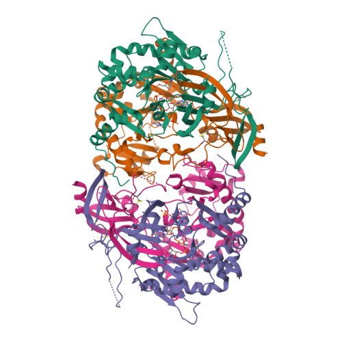
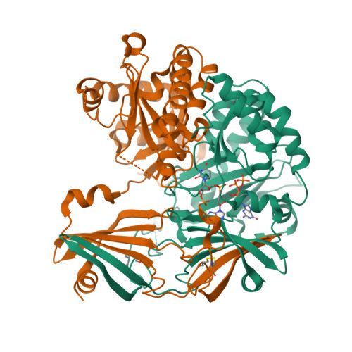
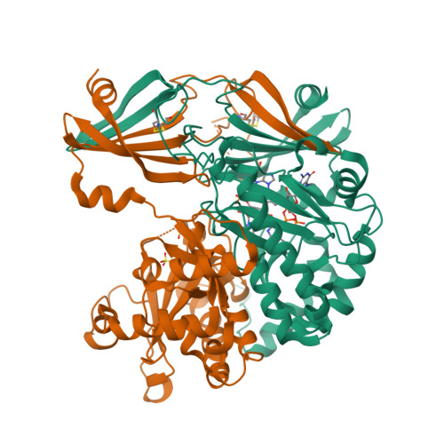
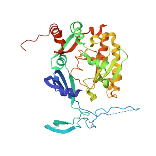
S.c. Scavenger Decapping Enzyme DcpS in complex with the capped RNA dinucleotide m7G-GU


Experimental Data Snapshot

  • Method: X-RAY DIFFRACTION
  • Resolution: 2.94 Å
  • R-Value Free: 0.263 
  • R-Value Work: 0.204 
  • R-Value Observed: 0.207 

Starting Model: experimental
View more details

wwPDB Validation   3D Report Full Report


Ligand Structure Quality Assessment 


This is version 2.0 of the entry. See complete history


Literature

Molecular basis of the selective processing of short mRNA substrates by the DcpS mRNA decapping enzyme.

Fuchs, A.L.Wurm, J.P.Neu, A.Sprangers, R.

(2020) Proc Natl Acad Sci U S A 117: 19237-19244

  • DOI: https://doi.org/10.1073/pnas.2009362117
  • Primary Citation of Related Structures:  
    6GBS, 6TRQ

  • PubMed Abstract: 

    The 5' messenger RNA (mRNA) cap structure enhances translation and protects the transcript against exonucleolytic degradation. During mRNA turnover, this cap is removed from the mRNA. This decapping step is catalyzed by the Scavenger Decapping Enzyme (DcpS), in case the mRNA has been exonucleolyticly shortened from the 3' end by the exosome complex. Here, we show that DcpS only processes mRNA fragments that are shorter than three nucleotides in length. Based on a combination of methyl transverse relaxation optimized (TROSY) NMR spectroscopy and X-ray crystallography, we established that the DcpS substrate length-sensing mechanism is based on steric clashes between the enzyme and the third nucleotide of a capped mRNA. For longer mRNA substrates, these clashes prevent conformational changes in DcpS that are required for the formation of a catalytically competent active site. Point mutations that enlarge the space for the third nucleotide in the mRNA body enhance the activity of DcpS on longer mRNA species. We find that this mechanism to ensure that the enzyme is not active on translating long mRNAs is conserved from yeast to humans. Finally, we show that the products that the exosome releases after 3' to 5' degradation of the mRNA body are indeed short enough to be decapped by DcpS. Our data thus directly confirms the notion that mRNA products of the exosome are direct substrates for DcpS. In summary, we demonstrate a direct relationship between conformational changes and enzyme activity that is exploited to achieve substrate selectivity.


  • Organizational Affiliation

    Department of Biophysics I, Regensburg Center for Biochemistry, University of Regensburg, 93053 Regensburg, Germany.


Macromolecules
Find similar proteins by:  (by identity cutoff)  |  3D Structure
Entity ID: 1
MoleculeChains Sequence LengthOrganismDetailsImage
m7GpppX diphosphatase
A, B, C, D
345Saccharomyces cerevisiae S288CMutation(s): 1 
Gene Names: DCS1YLR270W
EC: 3.6.1.59
UniProt
Find proteins for Q06151 (Saccharomyces cerevisiae (strain ATCC 204508 / S288c))
Go to UniProtKB:  Q06151
Sequence Annotations
Expand
  • Reference Sequence
Small Molecules
Ligands 5 Unique
IDChains Name / Formula / InChI Key2D Diagram3D Interactions
M7G
Query on M7G

Download Ideal Coordinates CCD File 
H [auth A],
L [auth C]
7N-METHYL-8-HYDROGUANOSINE-5'-DIPHOSPHATE
C11 H18 N5 O11 P2
SBASPRRECYVBRF-KQYNXXCUSA-O
0G
Query on 0G

Download Ideal Coordinates CCD File 
G [auth A],
K [auth C]
L-GUANOSINE-5'-MONOPHOSPHATE
C10 H14 N5 O8 P
RQFCJASXJCIDSX-GIMIYPNGSA-N
U
Query on U

Download Ideal Coordinates CCD File 
F [auth A],
I [auth C]
URIDINE-5'-MONOPHOSPHATE
C9 H13 N2 O9 P
DJJCXFVJDGTHFX-XVFCMESISA-N
SO4
Query on SO4

Download Ideal Coordinates CCD File 
M [auth D],
N [auth D]
SULFATE ION
O4 S
QAOWNCQODCNURD-UHFFFAOYSA-L
2PO
Query on 2PO

Download Ideal Coordinates CCD File 
E [auth A],
J [auth C]
PHOSPHONATE
H O3 P
ABLZXFCXXLZCGV-UHFFFAOYSA-L
Experimental Data & Validation

Experimental Data

  • Method: X-RAY DIFFRACTION
  • Resolution: 2.94 Å
  • R-Value Free: 0.263 
  • R-Value Work: 0.204 
  • R-Value Observed: 0.207 
  • Space Group: P 21 21 21
Unit Cell:
Length ( Å )Angle ( ˚ )
a = 87.761α = 90
b = 104.103β = 90
c = 189.223γ = 90
Software Package:
Software NamePurpose
PHENIXrefinement
XDSdata reduction
XSCALEdata scaling
PHASERphasing
PDB_EXTRACTdata extraction

Structure Validation

View Full Validation Report


Ligand Structure Quality Assessment 


Entry History & Funding Information

Deposition Data


Funding OrganizationLocationGrant Number
European Research CouncilGermany616052
German Research FoundationGermanySFB 960/2, B12

Revision History  (Full details and data files)

  • Version 1.0: 2020-07-22
    Type: Initial release
  • Version 1.1: 2020-08-12
    Changes: Database references, Derived calculations
  • Version 1.2: 2020-08-26
    Changes: Database references
  • Version 1.3: 2024-01-24
    Changes: Data collection, Database references, Refinement description
  • Version 2.0: 2024-03-06
    Changes: Atomic model, Data collection, Non-polymer description, Structure summary