6U1R

SxtG an amidinotransferase from the Microseira wollei in Saxitoxin biosynthetic pathway


Experimental Data Snapshot

  • Method: X-RAY DIFFRACTION
  • Resolution: 1.79 Å
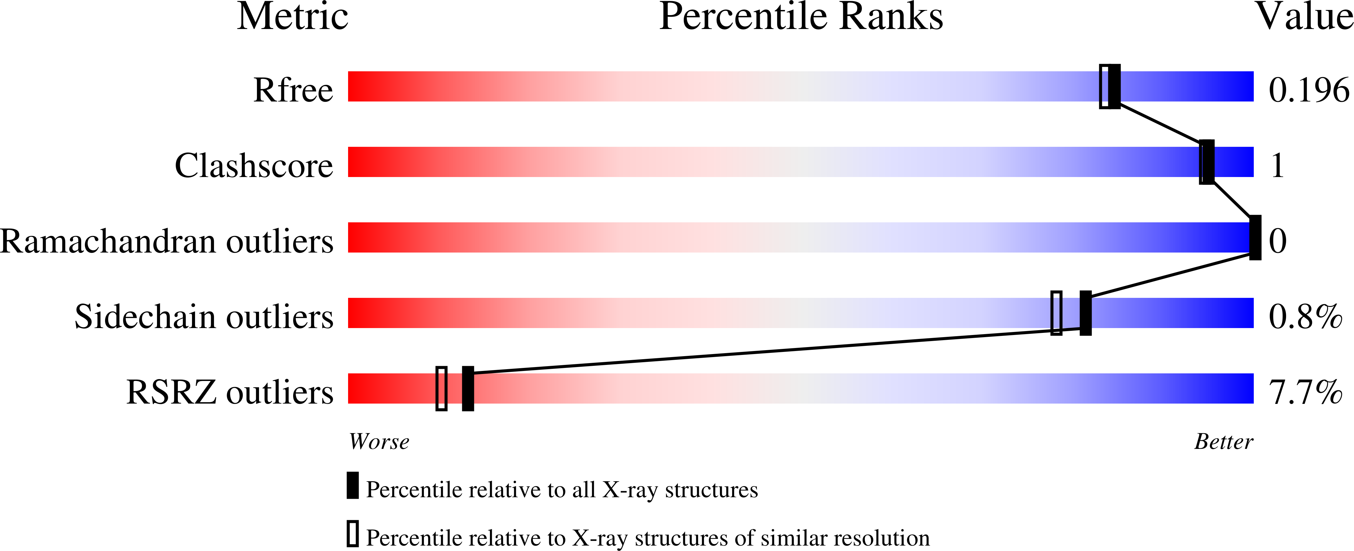
  • R-Value Free: 0.195 
  • R-Value Work: 0.167 
  • R-Value Observed: 0.169 

Starting Model: experimental
View more details

wwPDB Validation   3D Report Full Report


This is version 1.2 of the entry. See complete history


Literature

Substrate Promiscuity of a Paralytic Shellfish Toxin Amidinotransferase.

Lukowski, A.L.Mallik, L.Hinze, M.E.Carlson, B.M.Ellinwood, D.C.Pyser, J.B.Koutmos, M.Narayan, A.R.H.

(2020) ACS Chem Biol 15: 626-631

  • DOI: https://doi.org/10.1021/acschembio.9b00964
  • Primary Citation of Related Structures:  
    6U1R

  • PubMed Abstract: 

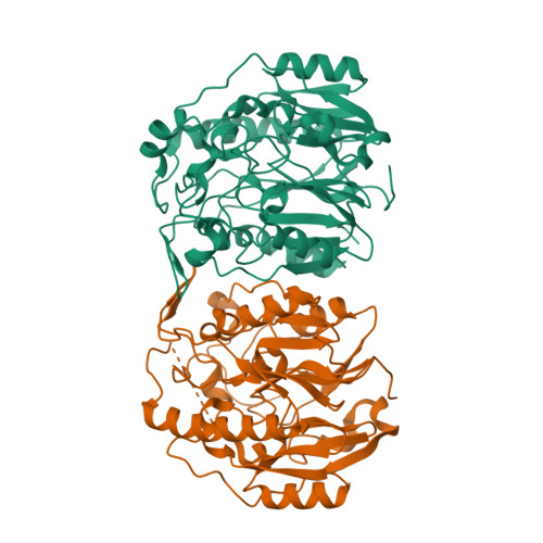
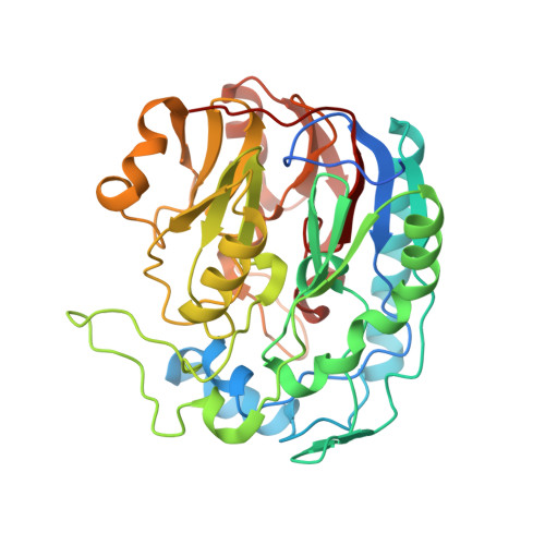
    Secondary metabolites are assembled by enzymes that often perform reactions with high selectivity and specificity. Many of these enzymes also tolerate variations in substrate structure, exhibiting promiscuity that enables various applications of a given biocatalyst. However, initial enzyme characterization studies frequently do not explore beyond the native substrates. This limited assessment of substrate scope contributes to the difficulty of identifying appropriate enzymes for specific synthetic applications. Here, we report the natural function of cyanobacterial SxtG, an amidinotransferase involved in the biosynthesis of paralytic shellfish toxins, and demonstrate its ability to modify a breadth of non-native substrates. In addition, we report the first X-ray crystal structure of SxtG, which provides rationale for this enzyme's substrate scope. Taken together, these data confirm the function of SxtG and exemplify its potential utility in biocatalytic synthesis.


Macromolecules
Find similar proteins by:  (by identity cutoff)  |  3D Structure
Entity ID: 1
MoleculeChains Sequence LengthOrganismDetailsImage
SxtG
A, B
383Microseira wolleiMutation(s): 0 
Gene Names: sxtG
UniProt
Find proteins for C3RVP0 (Microseira wollei)
Explore C3RVP0 
Go to UniProtKB:  C3RVP0
Entity Groups  
Sequence Clusters30% Identity50% Identity70% Identity90% Identity95% Identity100% Identity
UniProt GroupC3RVP0
Sequence Annotations
Expand
  • Reference Sequence
Small Molecules
Ligands 1 Unique
IDChains Name / Formula / InChI Key2D Diagram3D Interactions
FMT
Query on FMT

Download Ideal Coordinates CCD File 
C [auth A]FORMIC ACID
C H2 O2
BDAGIHXWWSANSR-UHFFFAOYSA-N
Experimental Data & Validation

Experimental Data

  • Method: X-RAY DIFFRACTION
  • Resolution: 1.79 Å
  • R-Value Free: 0.195 
  • R-Value Work: 0.167 
  • R-Value Observed: 0.169 
  • Space Group: P 2 21 21
Unit Cell:
Length ( Å )Angle ( ˚ )
a = 49.378α = 90
b = 100.562β = 90
c = 163.941γ = 90
Software Package:
Software NamePurpose
PHENIXrefinement
XDSdata reduction
Aimlessdata scaling
PHASERphasing

Structure Validation

View Full Validation Report


Entry History 

Deposition Data

Revision History  (Full details and data files)

  • Version 1.0: 2020-03-18
    Type: Initial release
  • Version 1.1: 2020-04-01
    Changes: Database references
  • Version 1.2: 2023-10-11
    Changes: Data collection, Database references, Refinement description