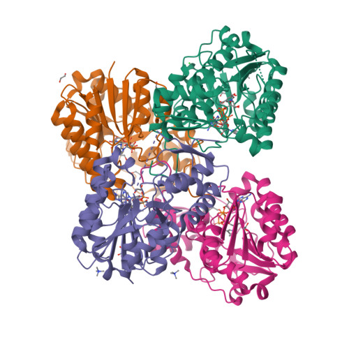
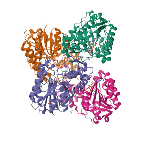
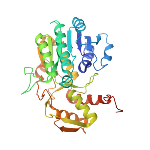
6VO6

Crystal Structure of Cj1427, an Essential NAD-dependent Dehydrogenase from Campylobacter jejuni, in the Presence of NADH and GDP


Experimental Data Snapshot

  • Method: X-RAY DIFFRACTION
  • Resolution: 1.50 Å
  • R-Value Free: 0.189 
  • R-Value Work: 0.163 
  • R-Value Observed: 0.164 

wwPDB Validation   3D Report Full Report


Ligand Structure Quality Assessment 


This is version 1.2 of the entry. See complete history


Literature

Structural Analysis of Cj1427, an Essential NAD-Dependent Dehydrogenase for the Biosynthesis of the Heptose Residues in the Capsular Polysaccharides ofCampylobacter jejuni.

Huddleston, J.P.Anderson, T.K.Spencer, K.D.Thoden, J.B.Raushel, F.M.Holden, H.M.

(2020) Biochemistry 59: 1314-1327

  • DOI: https://doi.org/10.1021/acs.biochem.0c00096
  • Primary Citation of Related Structures:  
    6VO6, 6VO8

  • PubMed Abstract: 

    Many strains of Campylobacter jejuni display modified heptose residues in their capsular polysaccharides (CPS). The precursor heptose was previously shown to be GDP-d- glycero -α-d- manno -heptose, from which a variety of modifications of the sugar moiety have been observed. These modifications include the generation of 6-deoxy derivatives and alterations of the stereochemistry at C3-C6. Previous work has focused on the enzymes responsible for the generation of the 6-deoxy derivatives and those involved in altering the stereochemistry at C3 and C5. However, the generation of the 6-hydroxyl heptose residues remains uncertain due to the lack of a specific enzyme to catalyze the initial oxidation at C4 of GDP-d- glycero -α-d- manno -heptose. Here we reexamine the previously reported role of Cj1427, a dehydrogenase found in C. jejuni NTCC 11168 (HS:2). We show that Cj1427 is co-purified with bound NADH, thus hindering catalysis of oxidation reactions. However, addition of a co-substrate, α-ketoglutarate, converts the bound NADH to NAD + . In this form, Cj1427 catalyzes the oxidation of l-2-hydroxyglutarate back to α-ketoglutarate. The crystal structure of Cj1427 with bound GDP-d- glycero -α-d- manno -heptose shows that the NAD(H) cofactor is ideally positioned to catalyze the oxidation at C4 of the sugar substrate. Additionally, the overall fold of the Cj1427 subunit places it into the well-defined short-chain dehydrogenase/reductase superfamily. The observed quaternary structure of the tetrameric enzyme, however, is highly unusual for members of this superfamily.


  • Organizational Affiliation

    Department of Chemistry, Texas A&M University, College Station, Texas 77843, United States.


Macromolecules
Find similar proteins by:  (by identity cutoff)  |  3D Structure
Entity ID: 1
MoleculeChains Sequence LengthOrganismDetailsImage
Putative sugar-nucleotide epimerase/dehydratease
A, B, C, D
321Campylobacter jejuni subsp. jejuni NCTC 11168 = ATCC 700819Mutation(s): 0 
Gene Names: Cj1427c
EC: 5.1.3.2 (PDB Primary Data), 1.1.98 (UniProt)
UniProt
Find proteins for Q0P8I7 (Campylobacter jejuni subsp. jejuni serotype O:2 (strain ATCC 700819 / NCTC 11168))
Explore Q0P8I7 
Go to UniProtKB:  Q0P8I7
Entity Groups  
Sequence Clusters30% Identity50% Identity70% Identity90% Identity95% Identity100% Identity
UniProt GroupQ0P8I7
Sequence Annotations
Expand
  • Reference Sequence
Small Molecules
Ligands 6 Unique
IDChains Name / Formula / InChI Key2D Diagram3D Interactions
NAI
Query on NAI

Download Ideal Coordinates CCD File 
E [auth A],
M [auth B],
T [auth C],
Y [auth D]
1,4-DIHYDRONICOTINAMIDE ADENINE DINUCLEOTIDE
C21 H29 N7 O14 P2
BOPGDPNILDQYTO-NNYOXOHSSA-N
GDP
Query on GDP

Download Ideal Coordinates CCD File 
F [auth A],
Z [auth D]
GUANOSINE-5'-DIPHOSPHATE
C10 H15 N5 O11 P2
QGWNDRXFNXRZMB-UUOKFMHZSA-N
TMA
Query on TMA

Download Ideal Coordinates CCD File 
DA [auth D],
L [auth A],
R [auth B],
S [auth B],
X [auth C]
TETRAMETHYLAMMONIUM ION
C4 H12 N
QEMXHQIAXOOASZ-UHFFFAOYSA-N
EDO
Query on EDO

Download Ideal Coordinates CCD File 
BA [auth D]
CA [auth D]
G [auth A]
H [auth A]
I [auth A]
BA [auth D],
CA [auth D],
G [auth A],
H [auth A],
I [auth A],
J [auth A],
N [auth B],
O [auth B],
P [auth B],
Q [auth B],
U [auth C],
V [auth C],
W [auth C]
1,2-ETHANEDIOL
C2 H6 O2
LYCAIKOWRPUZTN-UHFFFAOYSA-N
CL
Query on CL

Download Ideal Coordinates CCD File 
K [auth A]CHLORIDE ION
Cl
VEXZGXHMUGYJMC-UHFFFAOYSA-M
NA
Query on NA

Download Ideal Coordinates CCD File 
AA [auth D]SODIUM ION
Na
FKNQFGJONOIPTF-UHFFFAOYSA-N
Experimental Data & Validation

Experimental Data

  • Method: X-RAY DIFFRACTION
  • Resolution: 1.50 Å
  • R-Value Free: 0.189 
  • R-Value Work: 0.163 
  • R-Value Observed: 0.164 
  • Space Group: C 1 2 1
Unit Cell:
Length ( Å )Angle ( ˚ )
a = 132.608α = 90
b = 84.343β = 96.97
c = 135.45γ = 90
Software Package:
Software NamePurpose
REFMACrefinement
PDB_EXTRACTdata extraction
HKL-3000data reduction
HKL-3000data scaling
CRANKphasing

Structure Validation

View Full Validation Report


Ligand Structure Quality Assessment 


Entry History & Funding Information

Deposition Data


Funding OrganizationLocationGrant Number
National Institutes of Health/National Institute of General Medical Sciences (NIH/NIGMS)United StatesGM122825

Revision History  (Full details and data files)

  • Version 1.0: 2020-04-01
    Type: Initial release
  • Version 1.1: 2020-04-22
    Changes: Database references
  • Version 1.2: 2024-03-06
    Changes: Data collection, Database references