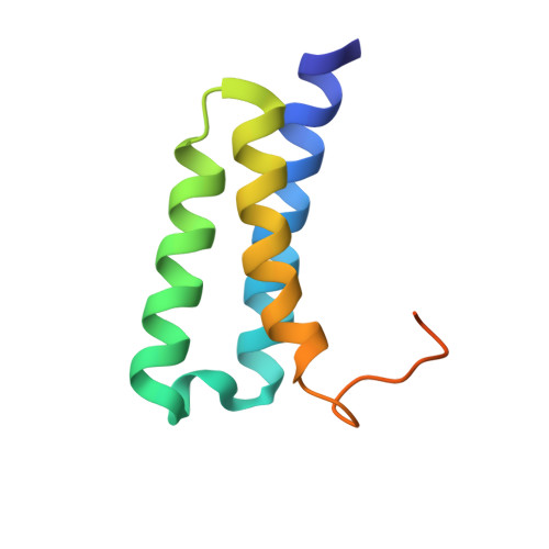
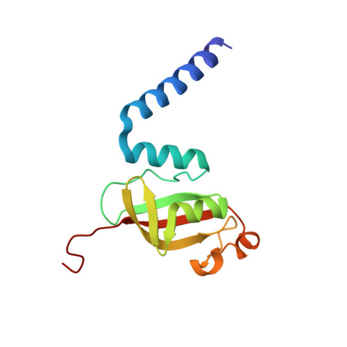
Transient and stabilized complexes of Nsp7, Nsp8, and Nsp12 in SARS-CoV-2 replication.
Wilamowski, M., Hammel, M., Leite, W., Zhang, Q., Kim, Y., Weiss, K.L., Jedrzejczak, R., Rosenberg, D.J., Fan, Y., Wower, J., Bierma, J.C., Sarker, A.H., Tsutakawa, S.E., Pingali, S.V., O'Neill, H.M., Joachimiak, A., Hura, G.L.(2021) Biophys J 120: 3152-3165
- PubMed: 34197805
- DOI: https://doi.org/10.1016/j.bpj.2021.06.006
- Primary Citation of Related Structures:
6WIQ, 6WQD, 6XIP - PubMed Abstract:
The replication transcription complex (RTC) from the virus SARS-CoV-2 is responsible for recognizing and processing RNA for two principal purposes. The RTC copies viral RNA for propagation into new virus and for ribosomal transcription of viral proteins. To accomplish these activities, the RTC mechanism must also conform to a large number of imperatives, including RNA over DNA base recognition, basepairing, distinguishing viral and host RNA, production of mRNA that conforms to host ribosome conventions, interfacing with error checking machinery, and evading host immune responses. In addition, the RTC will discontinuously transcribe specific sections of viral RNA to amplify certain proteins over others. Central to SARS-CoV-2 viability, the RTC is therefore dynamic and sophisticated. We have conducted a systematic structural investigation of three components that make up the RTC: Nsp7, Nsp8, and Nsp12 (also known as RNA-dependent RNA polymerase). We have solved high-resolution crystal structures of the Nsp7/8 complex, providing insight into the interaction between the proteins. We have used small-angle x-ray and neutron solution scattering (SAXS and SANS) on each component individually as pairs and higher-order complexes and with and without RNA. Using size exclusion chromatography and multiangle light scattering-coupled SAXS, we defined which combination of components forms transient or stable complexes. We used contrast-matching to mask specific complex-forming components to test whether components change conformation upon complexation. Altogether, we find that individual Nsp7, Nsp8, and Nsp12 structures vary based on whether other proteins in their complex are present. Combining our crystal structure, atomic coordinates reported elsewhere, SAXS, SANS, and other biophysical techniques, we provide greater insight into the RTC assembly, mechanism, and potential avenues for disruption of the complex and its functions.
Organizational Affiliation:
Center for Structural Genomics of Infectious Diseases, Consortium for Advanced Science and Engineering, University of Chicago, Chicago, Illinois; Department of Biochemistry and Molecular Biology, University of Chicago, Chicago, Illinois.


