6WYA

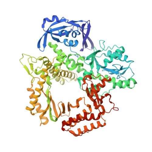
RTX (Reverse Transcription Xenopolymerase) in complex with a DNA duplex and dAMPNPP


Experimental Data Snapshot

  • Method: X-RAY DIFFRACTION
  • Resolution: 2.41 Å
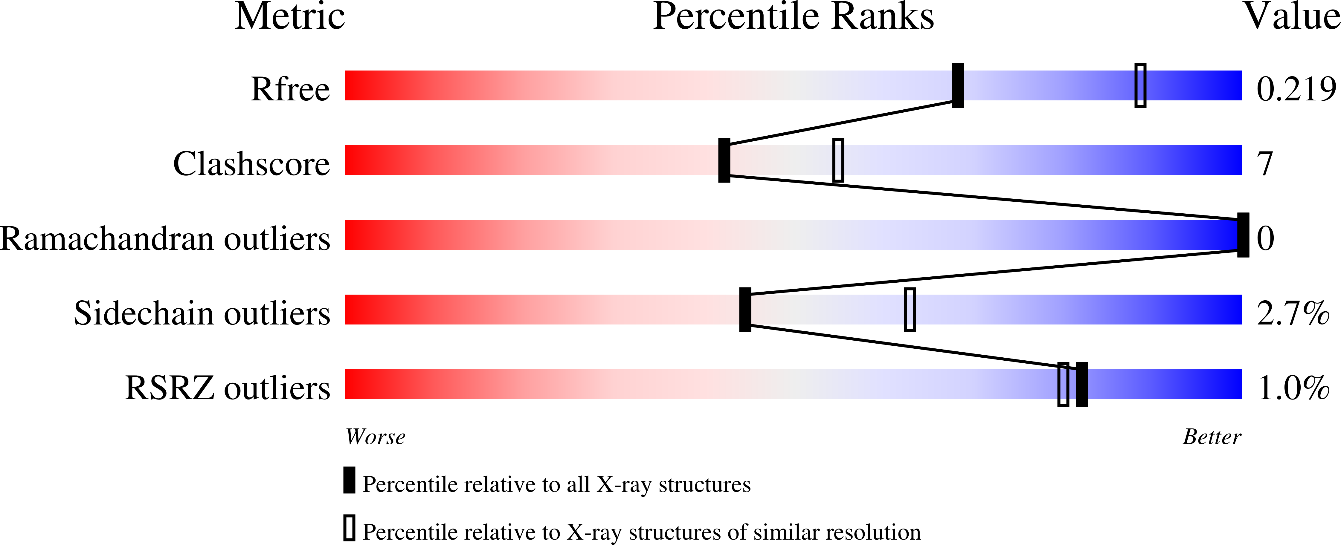
  • R-Value Free: 0.218 
  • R-Value Work: 0.166 
  • R-Value Observed: 0.167 

Starting Model: experimental
View more details

wwPDB Validation   3D Report Full Report


Ligand Structure Quality Assessment 


This is version 1.3 of the entry. See complete history


Literature

How a B family DNA polymerase has been evolved to copy RNA.

Choi, W.S.He, P.Pothukuchy, A.Gollihar, J.Ellington, A.D.Yang, W.

(2020) Proc Natl Acad Sci U S A 117: 21274-21280

  • DOI: https://doi.org/10.1073/pnas.2009415117
  • Primary Citation of Related Structures:  
    6WYA, 6WYB

  • PubMed Abstract: 

    We report here crystal structures of a reverse transcriptase RTX, which was evolved in vitro from the B family polymerase KOD, in complex with either a DNA duplex or an RNA-DNA hybrid. Compared with the apo, binary, and ternary complex structures of the original KOD polymerase, the 16 substitutions that result in the function of copying RNA to DNA do not change the overall protein structure. Only six substitutions occur at the substrate-binding surface, and the others change domain-domain interfaces in the polymerase to enable RNA-DNA hybrid binding and reverse transcription. Most notably, F587L at the Palm and Thumb interface stabilizes the open and apo conformation of the Thumb. The intrinsically flexible Thumb domain seems to play a major role in accommodating the RNA-DNA hybrid product distal to the active site. This is reminiscent of naturally occurring RNA-dependent DNA polymerases, including telomerase, which have a dramatically augmented Thumb domain, and of reverse transcriptase, which extends its Thumb with the RNase H domain.


  • Organizational Affiliation

    Laboratory of Molecular Biology, National Institute of Diabetes and Digestive and Kidney Diseases, NIH, Bethesda, MD 20892.


Macromolecules

Find similar proteins by:  (by identity cutoff)  |  3D Structure
Entity ID: 1
MoleculeChains Sequence LengthOrganismDetailsImage
DNA polymerase774Thermococcus kodakarensis KOD1Mutation(s): 0 
EC: 2.7.7.7
UniProt
Find proteins for D0VWU9 (Thermococcus kodakarensis (strain ATCC BAA-918 / JCM 12380 / KOD1))
Explore D0VWU9 
Go to UniProtKB:  D0VWU9
Entity Groups  
Sequence Clusters30% Identity50% Identity70% Identity90% Identity95% Identity100% Identity
UniProt GroupD0VWU9
Sequence Annotations
Expand
  • Reference Sequence

Find similar nucleic acids by:  Sequence   |   3D Structure  

Entity ID: 2
MoleculeChains LengthOrganismImage
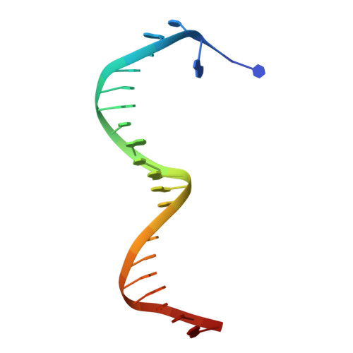
DNA strand 123Thermococcus kodakarensis KOD1
Sequence Annotations
Expand
  • Reference Sequence

Find similar nucleic acids by:  Sequence   |   3D Structure  

Entity ID: 3
MoleculeChains LengthOrganismImage
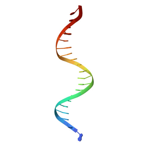
DNA strand 220Thermococcus kodakarensis KOD1
Sequence Annotations
Expand
  • Reference Sequence
Small Molecules
Ligands 5 Unique
IDChains Name / Formula / InChI Key2D Diagram3D Interactions
DZ4 (Subject of Investigation/LOI)
Query on DZ4

Download Ideal Coordinates CCD File 
H [auth A]2'-deoxy-5'-O-[(R)-hydroxy{[(R)-hydroxy(phosphonooxy)phosphoryl]amino}phosphoryl]adenosine
C10 H17 N6 O11 P3
WKIPJDSLGCBQCU-RRKCRQDMSA-N
TRS
Query on TRS

Download Ideal Coordinates CCD File 
I [auth A]2-AMINO-2-HYDROXYMETHYL-PROPANE-1,3-DIOL
C4 H12 N O3
LENZDBCJOHFCAS-UHFFFAOYSA-O
EDO
Query on EDO

Download Ideal Coordinates CCD File 
J [auth A],
M [auth B]
1,2-ETHANEDIOL
C2 H6 O2
LYCAIKOWRPUZTN-UHFFFAOYSA-N
ACT
Query on ACT

Download Ideal Coordinates CCD File 
K [auth A]ACETATE ION
C2 H3 O2
QTBSBXVTEAMEQO-UHFFFAOYSA-M
MG
Query on MG

Download Ideal Coordinates CCD File 
D [auth A],
E [auth A],
F [auth A],
G [auth A],
L [auth B]
MAGNESIUM ION
Mg
JLVVSXFLKOJNIY-UHFFFAOYSA-N
Experimental Data & Validation

Experimental Data

  • Method: X-RAY DIFFRACTION
  • Resolution: 2.41 Å
  • R-Value Free: 0.218 
  • R-Value Work: 0.166 
  • R-Value Observed: 0.167 
  • Space Group: P 21 21 21
Unit Cell:
Length ( Å )Angle ( ˚ )
a = 92.96α = 90
b = 96.36β = 90
c = 112.9γ = 90
Software Package:
Software NamePurpose
PHENIXrefinement
XDSdata reduction
XSCALEdata scaling
PHASERphasing

Structure Validation

View Full Validation Report


Ligand Structure Quality Assessment 


Entry History 

Deposition Data

Revision History  (Full details and data files)

  • Version 1.0: 2020-08-19
    Type: Initial release
  • Version 1.1: 2020-09-02
    Changes: Database references
  • Version 1.2: 2020-09-16
    Changes: Database references
  • Version 1.3: 2023-10-18
    Changes: Data collection, Database references, Refinement description