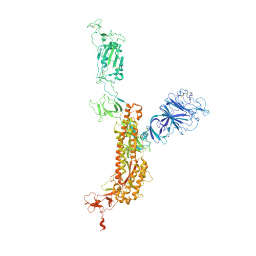
7MW5

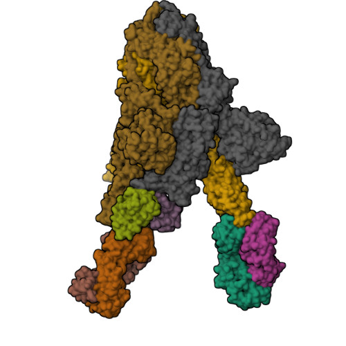
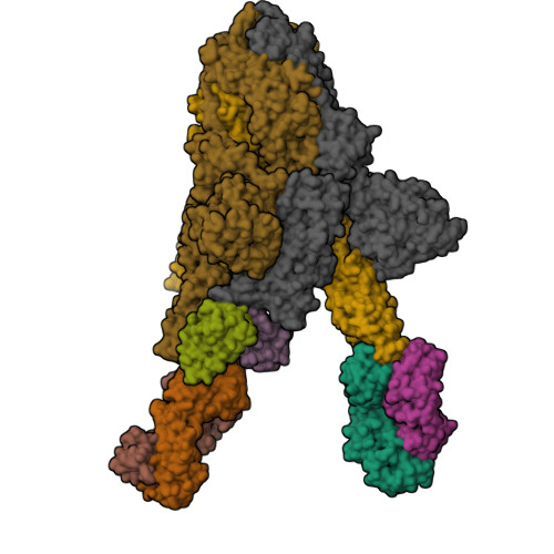
Structure of the SARS-CoV-2 Spike trimer with one RBD down in complex with the Fab fragment of human neutralizing antibody clone 2


Experimental Data Snapshot

  • Method: ELECTRON MICROSCOPY
  • Resolution: 3.42 Å
  • Aggregation State: PARTICLE 
  • Reconstruction Method: SINGLE PARTICLE 

wwPDB Validation   3D Report Full Report


This is version 1.2 of the entry. See complete history


Literature

Monospecific and bispecific monoclonal SARS-CoV-2 neutralizing antibodies that maintain potency against B.1.617.

Peng, L.Hu, Y.Mankowski, M.C.Ren, P.Chen, R.E.Wei, J.Zhao, M.Li, T.Tripler, T.Ye, L.Chow, R.D.Fang, Z.Wu, C.Dong, M.B.Cook, M.Wang, G.Clark, P.Nelson, B.Klein, D.Sutton, R.Diamond, M.S.Wilen, C.B.Xiong, Y.Chen, S.

(2022) Nat Commun 13: 1638-1638

  • DOI: https://doi.org/10.1038/s41467-022-29288-3
  • Primary Citation of Related Structures:  
    7MW2, 7MW3, 7MW4, 7MW5, 7MW6

  • PubMed Abstract: 

    COVID-19 pathogen SARS-CoV-2 has infected hundreds of millions and caused over 5 million deaths to date. Although multiple vaccines are available, breakthrough infections occur especially by emerging variants. Effective therapeutic options such as monoclonal antibodies (mAbs) are still critical. Here, we report the development, cryo-EM structures, and functional analyses of mAbs that potently neutralize SARS-CoV-2 variants of concern. By high-throughput single cell sequencing of B cells from spike receptor binding domain (RBD) immunized animals, we identify two highly potent SARS-CoV-2 neutralizing mAb clones that have single-digit nanomolar affinity and low-picomolar avidity, and generate a bispecific antibody. Lead antibodies show strong inhibitory activity against historical SARS-CoV-2 and several emerging variants of concern. We solve several cryo-EM structures at ~3 Å resolution of these neutralizing antibodies in complex with prefusion spike trimer ectodomain, and reveal distinct epitopes, binding patterns, and conformations. The lead clones also show potent efficacy in vivo against authentic SARS-CoV-2 in both prophylactic and therapeutic settings. We also generate and characterize a humanized antibody to facilitate translation and drug development. The humanized clone also has strong potency against both the original virus and the B.1.617.2 Delta variant. These mAbs expand the repertoire of therapeutics against SARS-CoV-2 and emerging variants.


  • Organizational Affiliation

    Department of Genetics, Yale University School of Medicine, New Haven, CT, USA.


Macromolecules
Find similar proteins by:  (by identity cutoff)  |  3D Structure
Entity ID: 1
MoleculeChains Sequence LengthOrganismDetailsImage
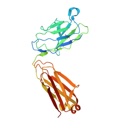
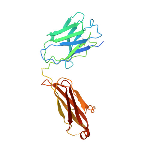
Fab of antibody clone 2, light chainA [auth L],
E [auth G],
H [auth E]
265Homo sapiensMutation(s): 0 
Entity Groups  
Sequence Clusters30% Identity50% Identity70% Identity90% Identity95% Identity100% Identity
Sequence Annotations
Expand
  • Reference Sequence
Find similar proteins by:  (by identity cutoff)  |  3D Structure
Entity ID: 2
MoleculeChains Sequence LengthOrganismDetailsImage
Fab of antibody clone 2, heavy chainB [auth H],
F,
I [auth D]
263Homo sapiensMutation(s): 0 
Entity Groups  
Sequence Clusters30% Identity50% Identity70% Identity90% Identity95% Identity100% Identity
Sequence Annotations
Expand
  • Reference Sequence
Find similar proteins by:  (by identity cutoff)  |  3D Structure
Entity ID: 3
MoleculeChains Sequence LengthOrganismDetailsImage
Spike glycoproteinC,
D [auth B],
G [auth A]
1,288Severe acute respiratory syndrome coronavirus 2Mutation(s): 0 
Gene Names: S2
UniProt
Find proteins for P0DTC2 (Severe acute respiratory syndrome coronavirus 2)
Explore P0DTC2 
Go to UniProtKB:  P0DTC2
Entity Groups  
Sequence Clusters30% Identity50% Identity70% Identity90% Identity95% Identity100% Identity
UniProt GroupP0DTC2
Glycosylation
Glycosylation Sites: 17Go to GlyGen: P0DTC2-1
Sequence Annotations
Expand
  • Reference Sequence
Oligosaccharides

Help

Entity ID: 4
MoleculeChains Length2D Diagram Glycosylation3D Interactions
2-acetamido-2-deoxy-beta-D-glucopyranose-(1-4)-2-acetamido-2-deoxy-beta-D-glucopyranose
AA [auth a],
BA [auth b],
CA [auth c],
DA [auth d],
EA [auth e],
AA [auth a],
BA [auth b],
CA [auth c],
DA [auth d],
EA [auth e],
FA [auth f],
GA [auth g],
HA [auth h],
IA [auth i],
J [auth I],
JA [auth j],
K [auth J],
KA [auth k],
L [auth K],
LA [auth l],
M,
N,
O,
P,
Q,
R,
S,
T,
U,
V,
W,
X,
Y,
Z
2N-Glycosylation
Glycosylation Resources
GlyTouCan:  G42666HT
GlyCosmos:  G42666HT
GlyGen:  G42666HT
Small Molecules
Ligands 1 Unique
IDChains Name / Formula / InChI Key2D Diagram3D Interactions
NAG (Subject of Investigation/LOI)
Query on NAG

Download Ideal Coordinates CCD File 
AB [auth B]
BB [auth B]
CB [auth B]
DB [auth A]
EB [auth A]
AB [auth B],
BB [auth B],
CB [auth B],
DB [auth A],
EB [auth A],
FB [auth A],
GB [auth A],
HB [auth A],
IB [auth A],
JB [auth A],
KB [auth A],
MA [auth C],
NA [auth C],
OA [auth C],
PA [auth C],
QA [auth C],
RA [auth C],
SA [auth C],
TA [auth C],
UA [auth B],
VA [auth B],
WA [auth B],
XA [auth B],
YA [auth B],
ZA [auth B]
2-acetamido-2-deoxy-beta-D-glucopyranose
C8 H15 N O6
OVRNDRQMDRJTHS-FMDGEEDCSA-N
Experimental Data & Validation

Experimental Data

  • Method: ELECTRON MICROSCOPY
  • Resolution: 3.42 Å
  • Aggregation State: PARTICLE 
  • Reconstruction Method: SINGLE PARTICLE 

Structure Validation

View Full Validation Report


Entry History & Funding Information

Deposition Data


Funding OrganizationLocationGrant Number
Department of Defense (DOD, United States)United States--

Revision History  (Full details and data files)

  • Version 1.0: 2022-03-16
    Type: Initial release
  • Version 1.1: 2023-11-01
    Changes: Data collection, Database references
  • Version 1.2: 2024-10-23
    Changes: Data collection, Structure summary