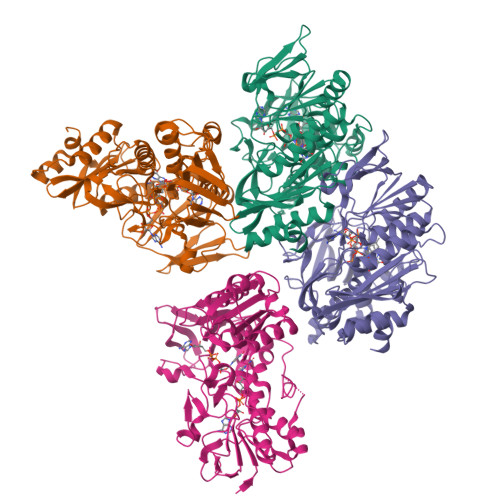
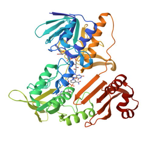
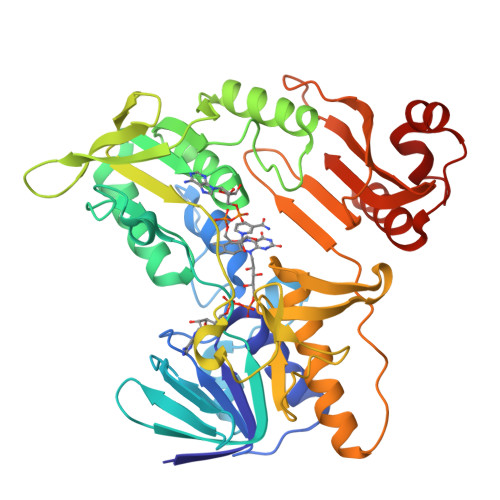
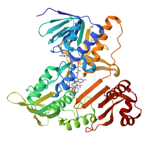
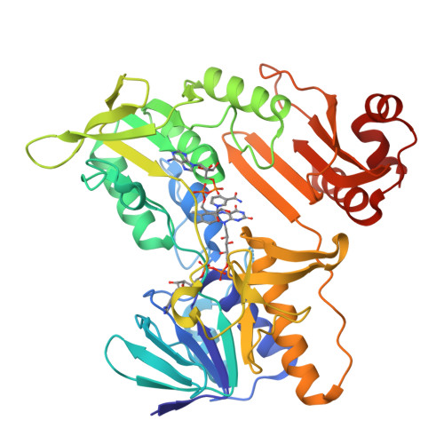
Crystal structure of Pennisetum glaucum monodehydroascorbate reductase
Sonkar, K.S., Arulandu, A., Achary, M.M., Reddy, M.K.To be published.
Experimental Data Snapshot
Starting Model: experimental
View more details
Entity ID: 1 | |||||
|---|---|---|---|---|---|
| Molecule | Chains | Sequence Length | Organism | Details | Image |
| Pennisetum glaucum monodehydroascorbate reductase | 435 | Cenchrus americanus | Mutation(s): 0 Gene Names: MDHAR EC: 1.6.5.4 | 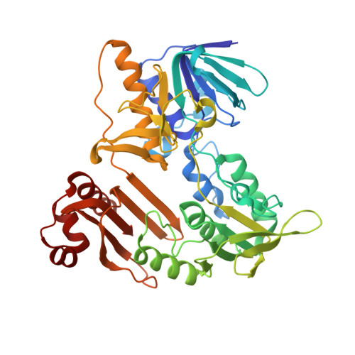 | |
Entity Groups | |||||
| Sequence Clusters | 30% Identity50% Identity70% Identity90% Identity95% Identity100% Identity | ||||
Sequence AnnotationsExpand | |||||
| |||||
| Ligands 2 Unique | |||||
|---|---|---|---|---|---|
| ID | Chains | Name / Formula / InChI Key | 2D Diagram | 3D Interactions | |
| FDA (Subject of Investigation/LOI) Query on FDA | F [auth A], H [auth B], J [auth C], L [auth D] | DIHYDROFLAVINE-ADENINE DINUCLEOTIDE C27 H35 N9 O15 P2 YPZRHBJKEMOYQH-UYBVJOGSSA-N |  | ||
| NAD Query on NAD | E [auth A], G [auth B], I [auth C], K [auth D] | NICOTINAMIDE-ADENINE-DINUCLEOTIDE C21 H27 N7 O14 P2 BAWFJGJZGIEFAR-NNYOXOHSSA-N |  | ||
| Length ( Å ) | Angle ( ˚ ) |
|---|---|
| a = 65.73 | α = 96.39 |
| b = 79.118 | β = 97.27 |
| c = 90.687 | γ = 111.92 |
| Software Name | Purpose |
|---|---|
| BUSTER | refinement |
| PDB_EXTRACT | data extraction |
| ARP/wARP | model building |
| BALBES | phasing |
| PHENIX | model building |
| autoPROC | data reduction |
| autoPROC | data scaling |
| Funding Organization | Location | Grant Number |
|---|---|---|
| Department of Biotechnology (DBT, India) | India | No.BT/PR18774/BIC/101/454/2016 |