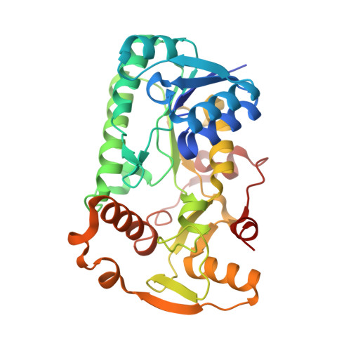
7CSF

AtPrR1 with NADP+ and (-)secoisolariciresinol


Experimental Data Snapshot

  • Method: X-RAY DIFFRACTION
  • Resolution: 1.98 Å
  • R-Value Free: 0.219 
  • R-Value Work: 0.178 
  • R-Value Observed: 0.179 

Starting Model: experimental
View more details

wwPDB Validation   3D Report Full Report


Ligand Structure Quality Assessment 


This is version 1.1 of the entry. See complete history


Literature

Structure-based engineering of substrate specificity for pinoresinol-lariciresinol reductases.

Xiao, Y.Shao, K.Zhou, J.Wang, L.Ma, X.Wu, D.Yang, Y.Chen, J.Feng, J.Qiu, S.Lv, Z.Zhang, L.Zhang, P.Chen, W.

(2021) Nat Commun 12: 2828-2828

  • DOI: https://doi.org/10.1038/s41467-021-23095-y
  • Primary Citation of Related Structures:  
    7CS2, 7CS3, 7CS4, 7CS5, 7CS6, 7CS7, 7CS8, 7CS9, 7CSA, 7CSB, 7CSC, 7CSD, 7CSE, 7CSF, 7CSG, 7CSH

  • PubMed Abstract: 

    Pinoresinol-lariciresinol reductases (PLRs) are enzymes involved in the lignan biosynthesis after the initial dimerization of two monolignols, and this represents the entry point for the synthesis of 8-8' lignans and contributes greatly to their structural diversity. Of particular interest has been the determination of how differing substrate specificities are achieved with these enzymes. Here, we present crystal structures of IiPLR1 from Isatis indigotica and pinoresinol reductases (PrRs) AtPrR1 and AtPrR2 from Arabidopsis thaliana, in the apo, substrate-bound and product-bound states. Each structure contains a head-to-tail homodimer, and the catalytic pocket comprises structural elements from both monomers. β4 loop covers the top of the pocket, and residue 98 from the loop governs catalytic specificity. The substrate specificities of IiPLR1 and AtPrR2 can be switched via structure-guided mutagenesis. Our study provides insight into the molecular mechanism underlying the substrate specificity of PLRs/PrRs and suggests an efficient strategy for the large-scale commercial production of the pharmaceutically valuable compound lariciresinol.


  • Organizational Affiliation

    Research and Development Center of Chinese Medicine Resources and Biotechnology, Institute of Chinese Materia Medica, Shanghai University of Traditional Chinese Medicine, Shanghai, China.


Macromolecules
Find similar proteins by:  (by identity cutoff)  |  3D Structure
Entity ID: 1
MoleculeChains Sequence LengthOrganismDetailsImage
Pinoresinol reductase 1A [auth C],
B [auth D],
C [auth A],
D [auth B]
317Arabidopsis thalianaMutation(s): 0 
Gene Names: PRR1PLR1At1g32100F3C3.10
EC: 1.23.1.1 (PDB Primary Data), 1.23.1.3 (PDB Primary Data)
UniProt
Find proteins for Q9FVQ6 (Arabidopsis thaliana)
Go to UniProtKB:  Q9FVQ6
Sequence Annotations
Expand
  • Reference Sequence
Small Molecules
Ligands 2 Unique
IDChains Name / Formula / InChI Key2D Diagram3D Interactions
NDP (Subject of Investigation/LOI)
Query on NDP

Download Ideal Coordinates CCD File 
F [auth C],
H [auth D],
J [auth A],
L [auth B]
NADPH DIHYDRO-NICOTINAMIDE-ADENINE-DINUCLEOTIDE PHOSPHATE
C21 H30 N7 O17 P3
ACFIXJIJDZMPPO-NNYOXOHSSA-N
GO6 (Subject of Investigation/LOI)
Query on GO6

Download Ideal Coordinates CCD File 
E [auth C],
G [auth D],
I [auth A],
K [auth B]
(2R,3R)-2,3-bis[(3-methoxy-4-oxidanyl-phenyl)methyl]butane-1,4-diol
C20 H26 O6
PUETUDUXMCLALY-HOTGVXAUSA-N
Experimental Data & Validation

Experimental Data

  • Method: X-RAY DIFFRACTION
  • Resolution: 1.98 Å
  • R-Value Free: 0.219 
  • R-Value Work: 0.178 
  • R-Value Observed: 0.179 
  • Space Group: P 1 21 1
Unit Cell:
Length ( Å )Angle ( ˚ )
a = 74.127α = 90
b = 143.273β = 116.376
c = 77.066γ = 90
Software Package:
Software NamePurpose
PHENIXrefinement
HKL-3000data reduction
HKL-3000data scaling
PHENIXphasing

Structure Validation

View Full Validation Report


Ligand Structure Quality Assessment 


Entry History 

Deposition Data

Revision History  (Full details and data files)

  • Version 1.0: 2021-06-09
    Type: Initial release
  • Version 1.1: 2023-11-29
    Changes: Data collection, Database references, Refinement description