7DZY

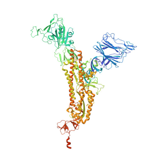
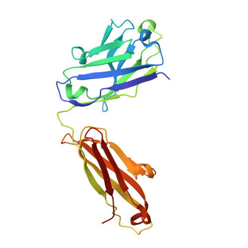
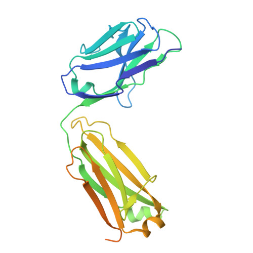
Spike protein from SARS-CoV2 with Fab fragment of enhancing antibody 2490


Experimental Data Snapshot

  • Method: ELECTRON MICROSCOPY
  • Resolution: 3.60 Å
  • Aggregation State: PARTICLE 
  • Reconstruction Method: SINGLE PARTICLE 

Starting Models: experimental
View more details

wwPDB Validation   3D Report Full Report


This is version 2.1 of the entry. See complete history


Literature

An infectivity-enhancing site on the SARS-CoV-2 spike protein targeted by antibodies.

Liu, Y.Soh, W.T.Kishikawa, J.I.Hirose, M.Nakayama, E.E.Li, S.Sasai, M.Suzuki, T.Tada, A.Arakawa, A.Matsuoka, S.Akamatsu, K.Matsuda, M.Ono, C.Torii, S.Kishida, K.Jin, H.Nakai, W.Arase, N.Nakagawa, A.Matsumoto, M.Nakazaki, Y.Shindo, Y.Kohyama, M.Tomii, K.Ohmura, K.Ohshima, S.Okamoto, T.Yamamoto, M.Nakagami, H.Matsuura, Y.Nakagawa, A.Kato, T.Okada, M.Standley, D.M.Shioda, T.Arase, H.

(2021) Cell 184: 3452-3466.e18

  • DOI: https://doi.org/10.1016/j.cell.2021.05.032
  • Primary Citation of Related Structures:  
    7DZW, 7DZX, 7DZY

  • PubMed Abstract: 

    Antibodies against the receptor-binding domain (RBD) of the SARS-CoV-2 spike protein prevent SARS-CoV-2 infection. However, the effects of antibodies against other spike protein domains are largely unknown. Here, we screened a series of anti-spike monoclonal antibodies from coronavirus disease 2019 (COVID-19) patients and found that some of antibodies against the N-terminal domain (NTD) induced the open conformation of RBD and thus enhanced the binding capacity of the spike protein to ACE2 and infectivity of SARS-CoV-2. Mutational analysis revealed that all of the infectivity-enhancing antibodies recognized a specific site on the NTD. Structural analysis demonstrated that all infectivity-enhancing antibodies bound to NTD in a similar manner. The antibodies against this infectivity-enhancing site were detected at high levels in severe patients. Moreover, we identified antibodies against the infectivity-enhancing site in uninfected donors, albeit at a lower frequency. These findings demonstrate that not only neutralizing antibodies but also enhancing antibodies are produced during SARS-CoV-2 infection.


  • Organizational Affiliation

    Department of Immunochemistry, Research Institute for Microbial Diseases, Osaka University, Osaka 565-0871, Japan; Laboratory of Immunochemistry, World Premier International Immunology Frontier Research Centre, Osaka University, Osaka 565-0871, Japan.


Macromolecules
Find similar proteins by:  (by identity cutoff)  |  3D Structure
Entity ID: 1
MoleculeChains Sequence LengthOrganismDetailsImage
Spike glycoprotein
A, B, C
1,249Severe acute respiratory syndrome coronavirus 2Mutation(s): 6 
Gene Names: S2
UniProt
Find proteins for P0DTC2 (Severe acute respiratory syndrome coronavirus 2)
Explore P0DTC2 
Go to UniProtKB:  P0DTC2
Entity Groups  
Sequence Clusters30% Identity50% Identity70% Identity90% Identity95% Identity100% Identity
UniProt GroupP0DTC2
Sequence Annotations
Expand
  • Reference Sequence
Find similar proteins by:  (by identity cutoff)  |  3D Structure
Entity ID: 2
MoleculeChains Sequence LengthOrganismDetailsImage
Fab Heavy chain of enhancing antibody 2490D [auth H],
F [auth M],
H [auth O]
225Homo sapiensMutation(s): 0 
Entity Groups  
Sequence Clusters30% Identity50% Identity70% Identity90% Identity95% Identity100% Identity
Sequence Annotations
Expand
  • Reference Sequence
Find similar proteins by:  (by identity cutoff)  |  3D Structure
Entity ID: 3
MoleculeChains Sequence LengthOrganismDetailsImage
Fab light chain of enhancing antibody 2490E [auth L],
G [auth N],
I [auth P]
266Homo sapiensMutation(s): 0 
Entity Groups  
Sequence Clusters30% Identity50% Identity70% Identity90% Identity95% Identity100% Identity
Sequence Annotations
Expand
  • Reference Sequence
Experimental Data & Validation

Experimental Data

  • Method: ELECTRON MICROSCOPY
  • Resolution: 3.60 Å
  • Aggregation State: PARTICLE 
  • Reconstruction Method: SINGLE PARTICLE 
EM Software:
TaskSoftware PackageVersion
RECONSTRUCTIONcryoSPARC3.0.1

Structure Validation

View Full Validation Report


Entry History & Funding Information

Deposition Data


Funding OrganizationLocationGrant Number
Japan Agency for Medical Research and Development (AMED)JapanJP19fk0108161
Japan Agency for Medical Research and Development (AMED)JapanJP20nf0101623
Japan Agency for Medical Research and Development (AMED)JapanJP20nk0101602
Japan Agency for Medical Research and Development (AMED)JapanJP17am0101108
Ministry of Education, Culture, Sports, Science and Technology (Japan)JapanJP19H04808
Japan Society for the Promotion of Science (JSPS)JapanJP18H05279
Japan Society for the Promotion of Science (JSPS)JapanJP19H03478

Revision History  (Full details and data files)

  • Version 1.0: 2021-06-02
    Type: Initial release
  • Version 1.1: 2021-07-07
    Changes: Database references
  • Version 2.0: 2023-08-02
    Type: Coordinate replacement
    Reason: Sequence discrepancy
    Changes: Advisory, Atomic model, Data collection, Database references, Derived calculations, Experimental preparation, Polymer sequence, Refinement description, Source and taxonomy, Structure summary
  • Version 2.1: 2024-06-05
    Changes: Data collection, Refinement description