7F6D

Reconstruction of the HerA-NurA complex from Deinococcus radiodurans


Experimental Data Snapshot

  • Method: ELECTRON MICROSCOPY
  • Resolution: 3.85 Å
  • Aggregation State: PARTICLE 
  • Reconstruction Method: SINGLE PARTICLE 

wwPDB Validation   3D Report Full Report


This is version 1.2 of the entry. See complete history


Literature

Mechanisms of helicase activated DNA end resection in bacteria.

Xu, Y.Xu, L.Qin, C.Wang, L.Guo, J.Hua, Y.Zhao, Y.

(2022) Structure 30: 1298-1306.e3

  • DOI: https://doi.org/10.1016/j.str.2022.06.005
  • Primary Citation of Related Structures:  
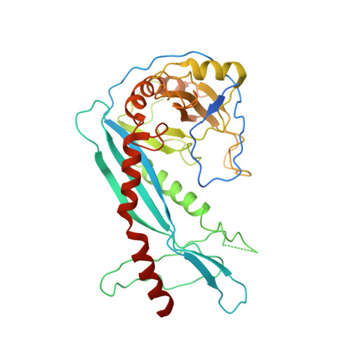
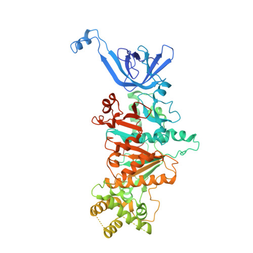
    7F6D

  • PubMed Abstract: 

    DNA end resection mediated by the coordinated action of nuclease and helicase is a crucial step in initiating homologous recombination. The end-resection apparatus NurA nuclease and HerA helicase are present in both archaea and bacteria. Here, we report the cryo-electron microscopy structure of a bacterial HerA-NurA complex from Deinococcus radiodurans. The structure reveals a barrel-like hexameric HerA and a distinctive NurA dimer subcomplex, which has a unique extended N-terminal region (ENR) involved in bacterial NurA dimerization and activation. In addition to the long protruding linking loop and the C-terminal α helix of NurA, the flexible ENR is close to the HerA-NurA interface and divides the central channel of the DrNurA dimer into two halves, suggesting a possible mechanism of DNA end processing. In summary, this work provides new insights into the structure, assembly, and activation mechanisms of bacterial DNA end resection mediated by a minimal end-resection apparatus.


  • Organizational Affiliation

    MOE Key Laboratory of Biosystems Homeostasis & Protection, Institute of Biophysics, College of Life Sciences, Zhejiang University, Hangzhou, China.


Macromolecules
Find similar proteins by:  (by identity cutoff)  |  3D Structure
Entity ID: 1
MoleculeChains Sequence LengthOrganismDetailsImage
NurAA [auth H],
B [auth G]
369Deinococcus radiodurans R1 = ATCC 13939 = DSM 20539Mutation(s): 0 
Gene Names: DR_0836
UniProt
Find proteins for Q9RW33 (Deinococcus radiodurans (strain ATCC 13939 / DSM 20539 / JCM 16871 / CCUG 27074 / LMG 4051 / NBRC 15346 / NCIMB 9279 / VKM B-1422 / R1))
Explore Q9RW33 
Go to UniProtKB:  Q9RW33
Entity Groups  
Sequence Clusters30% Identity50% Identity70% Identity90% Identity95% Identity100% Identity
UniProt GroupQ9RW33
Sequence Annotations
Expand
  • Reference Sequence
Find similar proteins by:  (by identity cutoff)  |  3D Structure
Entity ID: 2
MoleculeChains Sequence LengthOrganismDetailsImage
HerA618Deinococcus radiodurans R1 = ATCC 13939 = DSM 20539Mutation(s): 0 
Gene Names: DR_0837
UniProt
Find proteins for Q9RW32 (Deinococcus radiodurans (strain ATCC 13939 / DSM 20539 / JCM 16871 / CCUG 27074 / LMG 4051 / NBRC 15346 / NCIMB 9279 / VKM B-1422 / R1))
Explore Q9RW32 
Go to UniProtKB:  Q9RW32
Entity Groups  
Sequence Clusters30% Identity50% Identity70% Identity90% Identity95% Identity100% Identity
UniProt GroupQ9RW32
Sequence Annotations
Expand
  • Reference Sequence
Experimental Data & Validation

Experimental Data

  • Method: ELECTRON MICROSCOPY
  • Resolution: 3.85 Å
  • Aggregation State: PARTICLE 
  • Reconstruction Method: SINGLE PARTICLE 

Structure Validation

View Full Validation Report


Entry History & Funding Information

Deposition Data


Funding OrganizationLocationGrant Number
Ministry of Science and Technology (MoST, China)China2017YFA0503900

Revision History  (Full details and data files)

  • Version 1.0: 2022-06-29
    Type: Initial release
  • Version 1.1: 2022-08-17
    Changes: Database references
  • Version 1.2: 2022-09-14
    Changes: Database references