7JWB

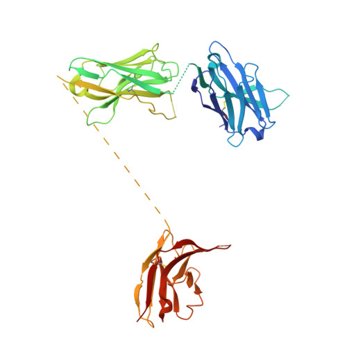
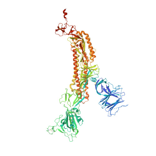
SARS CoV2 Spike ectodomain with engineered trimerized VH binder


Experimental Data Snapshot

  • Method: ELECTRON MICROSCOPY
  • Resolution: 6.00 Å
  • Resolution: 3.20 Å
  • Aggregation State: PARTICLE 
  • Reconstruction Method: SINGLE PARTICLE 

Starting Models: experimental
View more details

wwPDB Validation   3D Report Full Report


This is version 1.4 of the entry. See complete history


Literature

Bi-paratopic and multivalent VH domains block ACE2 binding and neutralize SARS-CoV-2.

Bracken, C.J.Lim, S.A.Solomon, P.Rettko, N.J.Nguyen, D.P.Zha, B.S.Schaefer, K.Byrnes, J.R.Zhou, J.Lui, I.Liu, J.Pance, K.Zhou, X.X.Leung, K.K.Wells, J.A.

(2021) Nat Chem Biol 17: 113-121

  • DOI: https://doi.org/10.1038/s41589-020-00679-1
  • Primary Citation of Related Structures:  
    7JWB

  • PubMed Abstract: 

    Neutralizing agents against SARS-CoV-2 are urgently needed for the treatment and prophylaxis of COVID-19. Here, we present a strategy to rapidly identify and assemble synthetic human variable heavy (VH) domains toward neutralizing epitopes. We constructed a VH-phage library and targeted the angiotensin-converting enzyme 2 (ACE2) binding interface of the SARS-CoV-2 Spike receptor-binding domain (Spike-RBD). Using a masked selection approach, we identified VH binders to two non-overlapping epitopes and further assembled these into multivalent and bi-paratopic formats. These VH constructs showed increased affinity to Spike (up to 600-fold) and neutralization potency (up to 1,400-fold) on pseudotyped SARS-CoV-2 virus when compared to standalone VH domains. The most potent binder, a trivalent VH, neutralized authentic SARS-CoV-2 with a half-maximal inhibitory concentration (IC 50 ) of 4.0 nM (180 ng ml -1 ). A cryo-EM structure of the trivalent VH bound to Spike shows each VH domain engaging an RBD at the ACE2 binding site, confirming our original design strategy.


  • Organizational Affiliation

    Department of Pharmaceutical Chemistry, University of California, San Francisco, San Francisco, CA, USA.


Macromolecules
Find similar proteins by:  (by identity cutoff)  |  3D Structure
Entity ID: 1
MoleculeChains Sequence LengthOrganismDetailsImage
autonomous human heavy chain variable domainA [auth D]421Homo sapiensMutation(s): 0 
Entity Groups  
Sequence Clusters30% Identity50% Identity70% Identity90% Identity95% Identity100% Identity
Sequence Annotations
Expand
  • Reference Sequence
Find similar proteins by:  (by identity cutoff)  |  3D Structure
Entity ID: 2
MoleculeChains Sequence LengthOrganismDetailsImage
Spike glycoproteinB [auth A],
C [auth B],
D [auth C]
1,208Severe acute respiratory syndrome coronavirus 2Mutation(s): 0 
Gene Names: S2
UniProt
Find proteins for P0DTC2 (Severe acute respiratory syndrome coronavirus 2)
Explore P0DTC2 
Go to UniProtKB:  P0DTC2
Entity Groups  
Sequence Clusters30% Identity50% Identity70% Identity90% Identity95% Identity100% Identity
UniProt GroupP0DTC2
Sequence Annotations
Expand
  • Reference Sequence
Experimental Data & Validation

Experimental Data

  • Method: ELECTRON MICROSCOPY
  • Resolution: 6.00 Å
  • Resolution: 3.20 Å
  • Aggregation State: PARTICLE 
  • Reconstruction Method: SINGLE PARTICLE 

Structure Validation

View Full Validation Report


Entry History & Funding Information

Deposition Data


Funding OrganizationLocationGrant Number
Defense Advanced Research Projects Agency (DARPA)United StatesHR0011-19-2-0020
Quantitative Biosciences InstituteUnited States--
FastGrantsUnited States--
Laboratory for Genomics Research (LGR)United StatesExcellence in Research Award (ERA)

Revision History  (Full details and data files)

  • Version 1.0: 2020-10-07
    Type: Initial release
  • Version 1.1: 2020-10-14
    Changes: Database references
  • Version 1.2: 2020-12-16
    Changes: Database references
  • Version 1.3: 2020-12-30
    Changes: Database references
  • Version 1.4: 2024-11-13
    Changes: Data collection, Database references, Refinement description, Structure summary