7KIP

A 3.4 Angstrom cryo-EM structure of the human coronavirus spike trimer computationally derived from vitrified NL63 virus particles


Experimental Data Snapshot

  • Method: ELECTRON MICROSCOPY
  • Resolution: 3.39 Å
  • Aggregation State: PARTICLE 
  • Reconstruction Method: SINGLE PARTICLE 

wwPDB Validation   3D Report Full Report


This is version 1.0 of the entry. See complete history

Re-refinement Note

A newer entry is available that reflects an alternative modeling of the original data: 8FR7


Literature

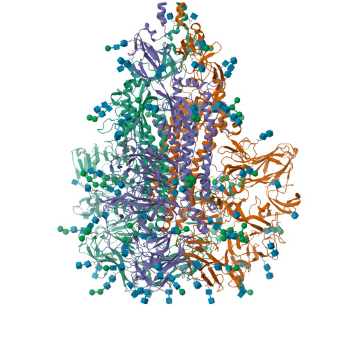
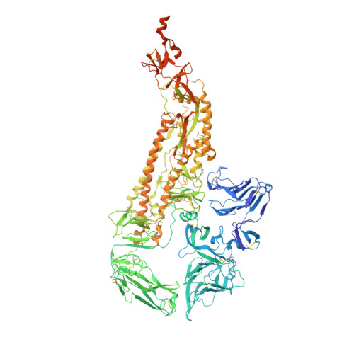
A 3.4- angstrom cryo-EM structure of the human coronavirus spike trimer computationally derived from vitrified NL63 virus particles.

Zhang, K.Li, S.Pintilie, G.Chmielewski, D.Schmid, M.F.Simmons, G.Jin, J.Chiu, W.

(2020) Biorxiv 

  • DOI: https://doi.org/10.1101/2020.08.11.245696
  • Primary Citation of Related Structures:  
    7KIP

  • PubMed Abstract: 

    Human coronavirus NL63 (HCoV-NL63) is an enveloped pathogen of the family Coronaviridae that spreads worldwide and causes up to 10% of all annual respiratory diseases. HCoV-NL63 is typically associated with mild upper respiratory symptoms in children, elderly and immunocompromised individuals. It has also been shown to cause severe lower respiratory illness. NL63 shares ACE2 as a receptor for viral entry with SARS-CoV and SARS-CoV-2. Here we present the in situ structure of HCoV-NL63 spike (S) trimer at 3.4-Å resolution by single-particle cryo-EM imaging of vitrified virions without chemical fixative. It is structurally homologous to that obtained previously from the biochemically purified ectodomain of HCoV-NL63 S trimer, which displays a 3-fold symmetric trimer in a single conformation. In addition to previously proposed and observed glycosylation sites, our map shows density at other amino acid positions as well as differences in glycan structures. The domain arrangement within a protomer is strikingly different from that of the SARS-CoV-2 S and may explain their different requirements for activating binding to the receptor. This structure provides the basis for future studies of spike proteins with receptors, antibodies, or drugs, in the native state of the coronavirus particles.


Macromolecules
Find similar proteins by:  (by identity cutoff)  |  3D Structure
Entity ID: 1
MoleculeChains Sequence LengthOrganismDetailsImage
Spike glycoprotein
A, B, C
1,356Human coronavirus NL63Mutation(s): 0 
UniProt
Find proteins for Q6Q1S2 (Human coronavirus NL63)
Explore Q6Q1S2 
Go to UniProtKB:  Q6Q1S2
Entity Groups  
Sequence Clusters30% Identity50% Identity70% Identity90% Identity95% Identity100% Identity
UniProt GroupQ6Q1S2
Sequence Annotations
Expand
  • Reference Sequence
Oligosaccharides

Help

Entity ID: 2
MoleculeChains Length2D Diagram Glycosylation3D Interactions
alpha-D-mannopyranose-(1-2)-alpha-D-mannopyranose-(1-3)-[alpha-D-mannopyranose-(1-6)]beta-D-mannopyranose-(1-4)-2-acetamido-2-deoxy-beta-D-glucopyranose-(1-4)-2-acetamido-2-deoxy-beta-D-glucopyranoseD,
TA [auth t],
Y
6N-Glycosylation
Glycosylation Resources
GlyTouCan:  G56014GC
GlyCosmos:  G56014GC
GlyGen:  G56014GC
Entity ID: 3
MoleculeChains Length2D Diagram Glycosylation3D Interactions
2-acetamido-2-deoxy-beta-D-glucopyranose-(1-4)-2-acetamido-2-deoxy-beta-D-glucopyranose
BA [auth b],
CB [auth 2],
DA [auth d],
E,
EA [auth e],
BA [auth b],
CB [auth 2],
DA [auth d],
E,
EA [auth e],
FB [auth 5],
G,
GB [auth 6],
HA [auth h],
HB [auth 7],
I,
IB [auth 8],
J,
JB [auth 9],
KA [auth k],
LA [auth l],
M,
MA [auth m],
MB [auth CA],
NA [auth n],
OA [auth o],
P,
Q,
R,
RA [auth r],
S,
T,
UA [auth u],
W,
WA [auth w],
YA [auth y],
Z,
ZA [auth z]
2N-Glycosylation
Glycosylation Resources
GlyTouCan:  G42666HT
GlyCosmos:  G42666HT
GlyGen:  G42666HT
Entity ID: 4
MoleculeChains Length2D Diagram Glycosylation3D Interactions
alpha-D-mannopyranose-(1-3)-beta-D-mannopyranose-(1-4)-2-acetamido-2-deoxy-beta-D-glucopyranose-(1-4)-2-acetamido-2-deoxy-beta-D-glucopyranose
AA [auth a],
F,
KB [auth AA],
PA [auth p],
U,
AA [auth a],
F,
KB [auth AA],
PA [auth p],
U,
VA [auth v]
4N-Glycosylation
Glycosylation Resources
GlyTouCan:  G81315DD
GlyCosmos:  G81315DD
GlyGen:  G81315DD
Entity ID: 5
MoleculeChains Length2D Diagram Glycosylation3D Interactions
alpha-D-mannopyranose-(1-6)-beta-D-mannopyranose-(1-4)-2-acetamido-2-deoxy-beta-D-glucopyranose-(1-4)-2-acetamido-2-deoxy-beta-D-glucopyranoseCA [auth c],
H,
XA [auth x]
4N-Glycosylation
Glycosylation Resources
GlyTouCan:  G22573RC
GlyCosmos:  G22573RC
GlyGen:  G22573RC
Entity ID: 6
MoleculeChains Length2D Diagram Glycosylation3D Interactions
alpha-D-mannopyranose-(1-2)-alpha-D-mannopyranose-(1-2)-alpha-D-mannopyranose-(1-3)-[alpha-D-mannopyranose-(1-3)-alpha-D-mannopyranose-(1-6)]beta-D-mannopyranose-(1-4)-2-acetamido-2-deoxy-beta-D-glucopyranose-(1-4)-2-acetamido-2-deoxy-beta-D-glucopyranoseAB [auth 0],
FA [auth f],
K
8N-Glycosylation
Glycosylation Resources
GlyTouCan:  G83582BK
GlyCosmos:  G83582BK
GlyGen:  G83582BK
Entity ID: 7
MoleculeChains Length2D Diagram Glycosylation3D Interactions
beta-D-mannopyranose-(1-4)-2-acetamido-2-deoxy-beta-D-glucopyranoseBB [auth 1],
GA [auth g],
L
2N/A
Glycosylation Resources
GlyTouCan:  G90333CG
GlyCosmos:  G90333CG
GlyGen:  G90333CG
Entity ID: 8
MoleculeChains Length2D Diagram Glycosylation3D Interactions
alpha-D-mannopyranose-(1-3)-[alpha-D-mannopyranose-(1-6)]beta-D-mannopyranose-(1-4)-2-acetamido-2-deoxy-beta-D-glucopyranose-(1-4)-2-acetamido-2-deoxy-beta-D-glucopyranose
DB [auth 3],
IA [auth i],
LB [auth BA],
N,
QA [auth q],
DB [auth 3],
IA [auth i],
LB [auth BA],
N,
QA [auth q],
V
5N-Glycosylation
Glycosylation Resources
GlyTouCan:  G22768VO
GlyCosmos:  G22768VO
GlyGen:  G22768VO
Entity ID: 9
MoleculeChains Length2D Diagram Glycosylation3D Interactions
beta-D-mannopyranose-(1-4)-2-acetamido-2-deoxy-beta-D-glucopyranose-(1-4)-2-acetamido-2-deoxy-beta-D-glucopyranose
EB [auth 4],
JA [auth j],
NB [auth DA],
O,
SA [auth s],
EB [auth 4],
JA [auth j],
NB [auth DA],
O,
SA [auth s],
X
3N-Glycosylation
Glycosylation Resources
GlyTouCan:  G15407YE
GlyCosmos:  G15407YE
GlyGen:  G15407YE
Small Molecules
Ligands 1 Unique
IDChains Name / Formula / InChI Key2D Diagram3D Interactions
NAG (Subject of Investigation/LOI)
Query on NAG

Download Ideal Coordinates CCD File 
AC [auth B]
BC [auth B]
CC [auth B]
DC [auth B]
EC [auth C]
AC [auth B],
BC [auth B],
CC [auth B],
DC [auth B],
EC [auth C],
FC [auth C],
GC [auth C],
HC [auth C],
IC [auth C],
JC [auth C],
KC [auth C],
LC [auth C],
OB [auth A],
PB [auth A],
QB [auth A],
RB [auth A],
SB [auth A],
TB [auth A],
UB [auth A],
VB [auth A],
WB [auth B],
XB [auth B],
YB [auth B],
ZB [auth B]
2-acetamido-2-deoxy-beta-D-glucopyranose
C8 H15 N O6
OVRNDRQMDRJTHS-FMDGEEDCSA-N
Experimental Data & Validation

Experimental Data

  • Method: ELECTRON MICROSCOPY
  • Resolution: 3.39 Å
  • Aggregation State: PARTICLE 
  • Reconstruction Method: SINGLE PARTICLE 
EM Software:
TaskSoftware PackageVersion
RECONSTRUCTIONcryoSPARC2
MODEL REFINEMENTPHENIX

Structure Validation

View Full Validation Report


Entry History & Funding Information

Deposition Data


Funding OrganizationLocationGrant Number
National Institutes of Health/National Institute of General Medical Sciences (NIH/NIGMS)United StatesU24GM129564
National Institutes of Health/National Institute of General Medical Sciences (NIH/NIGMS)United StatesR01GM079429
National Institutes of Health/National Institute of General Medical Sciences (NIH/NIGMS)United StatesP41GM103832

Revision History  (Full details and data files)

  • Version 1.0: 2020-11-11
    Type: Initial release