7L4Z

Structure of SARS-CoV-2 spike RBD in complex with cyclic peptide


Experimental Data Snapshot

  • Method: X-RAY DIFFRACTION
  • Resolution: 3.96 Å
  • R-Value Free: 0.282 
  • R-Value Work: 0.257 
  • R-Value Observed: 0.258 

wwPDB Validation   3D Report Full Report


Ligand Structure Quality Assessment 


This is version 1.2 of the entry. See complete history


Literature

Discovery of Cyclic Peptide Ligands to the SARS-CoV-2 Spike Protein Using mRNA Display.

Norman, A.Franck, C.Christie, M.Hawkins, P.M.E.Patel, K.Ashhurst, A.S.Aggarwal, A.Low, J.K.K.Siddiquee, R.Ashley, C.L.Steain, M.Triccas, J.A.Turville, S.Mackay, J.P.Passioura, T.Payne, R.J.

(2021) ACS Cent Sci 7: 1001-1008

  • DOI: https://doi.org/10.1021/acscentsci.0c01708
  • Primary Citation of Related Structures:  
    7L4Z

  • PubMed Abstract: 

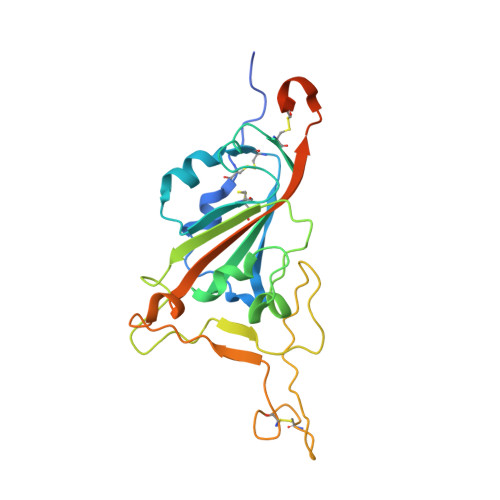
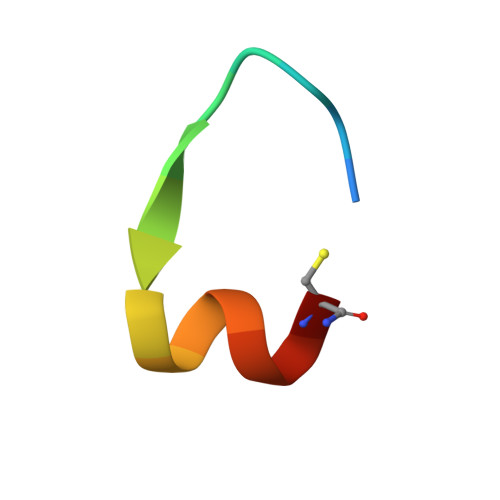
    The COVID-19 pandemic, caused by SARS-CoV-2, has led to substantial morbidity, mortality, and disruption globally. Cellular entry of SARS-CoV-2 is mediated by the viral spike protein, and affinity ligands to this surface protein have the potential for applications as antivirals and diagnostic reagents. Here, we describe the affinity selection of cyclic peptide ligands to the SARS-CoV-2 spike protein receptor binding domain (RBD) from three distinct libraries (in excess of a trillion molecules each) by mRNA display. We identified six high affinity molecules with dissociation constants ( K D ) in the nanomolar range (15-550 nM) to the RBD. The highest affinity ligand could be used as an affinity reagent to detect the spike protein in solution by ELISA, and the cocrystal structure of this molecule bound to the RBD demonstrated that it binds to a cryptic binding site, displacing a β-strand near the C-terminus. Our findings provide key mechanistic insight into the binding of peptide ligands to the SARS-CoV-2 spike RBD, and the ligands discovered in this work may find future use as reagents for diagnostic applications.


  • Organizational Affiliation

    School of Chemistry, The University of Sydney, Sydney, New South Wales 2006, Australia.


Macromolecules
Find similar proteins by:  (by identity cutoff)  |  3D Structure
Entity ID: 1
MoleculeChains Sequence LengthOrganismDetailsImage
Spike protein S1A [auth B],
B [auth D],
C [auth A],
D [auth C],
E
229Severe acute respiratory syndrome coronavirus 2Mutation(s): 0 
Gene Names: S2
UniProt
Find proteins for P0DTC2 (Severe acute respiratory syndrome coronavirus 2)
Explore P0DTC2 
Go to UniProtKB:  P0DTC2
Entity Groups  
Sequence Clusters30% Identity50% Identity70% Identity90% Identity95% Identity100% Identity
UniProt GroupP0DTC2
Sequence Annotations
Expand
  • Reference Sequence

Find similar proteins by:  Sequence   |   3D Structure  

Entity ID: 2
MoleculeChains Sequence LengthOrganismDetailsImage
ACE-DTY-LYS-ALA-GLY-VAL-VAL-TYR-GLY-TYR-ASN-ALA-TRP-ILE-ARG-CYS-NH2F [auth S],
G [auth T],
H [auth R],
I [auth U],
J [auth V]
17synthetic constructMutation(s): 0 
Entity Groups  
Sequence Clusters30% Identity50% Identity70% Identity90% Identity95% Identity100% Identity
Sequence Annotations
Expand
  • Reference Sequence
Experimental Data & Validation

Experimental Data

  • Method: X-RAY DIFFRACTION
  • Resolution: 3.96 Å
  • R-Value Free: 0.282 
  • R-Value Work: 0.257 
  • R-Value Observed: 0.258 
  • Space Group: H 3 2
Unit Cell:
Length ( Å )Angle ( ˚ )
a = 284.92α = 90
b = 284.92β = 90
c = 156.016γ = 120
Software Package:
Software NamePurpose
PHENIXrefinement
XDSdata reduction
Aimlessdata scaling
PHASERphasing

Structure Validation

View Full Validation Report


Ligand Structure Quality Assessment 


Entry History & Funding Information

Deposition Data


Funding OrganizationLocationGrant Number
National Health and Medical Research Council (NHMRC, Australia)AustraliaAPP1194941

Revision History  (Full details and data files)

  • Version 1.0: 2021-06-30
    Type: Initial release
  • Version 1.1: 2021-08-04
    Changes: Database references
  • Version 1.2: 2023-10-18
    Changes: Data collection, Database references, Refinement description