7LZU

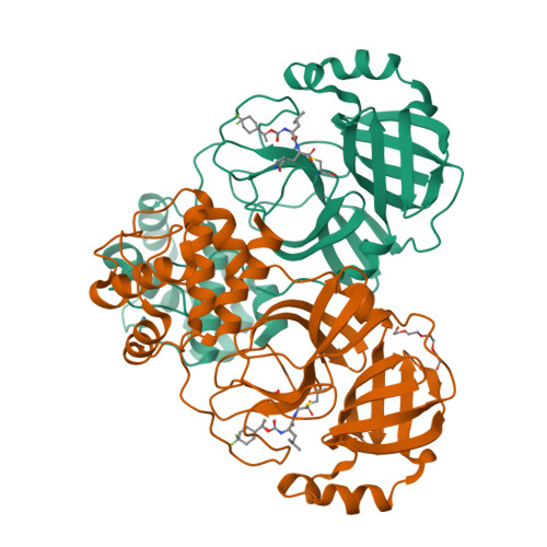
Structure of SARS-CoV-2 3CL protease in complex with inhibitor 12b


Experimental Data Snapshot

  • Method: X-RAY DIFFRACTION
  • Resolution: 1.60 Å
  • R-Value Free: 0.201 
  • R-Value Work: 0.178 
  • R-Value Observed: 0.179 

Starting Model: experimental
View more details

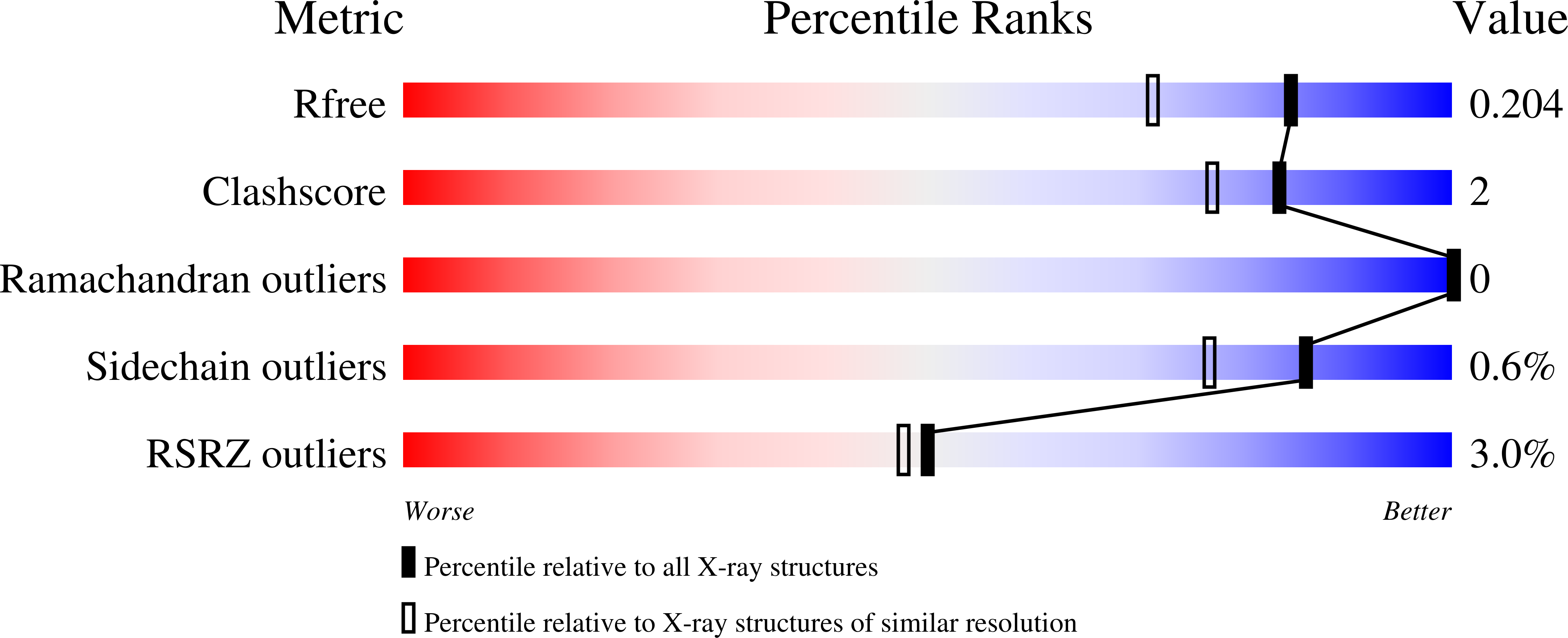
wwPDB Validation   3D Report Full Report


Ligand Structure Quality Assessment 


This is version 1.4 of the entry. See complete history


Literature

Structure-Guided Design of Potent Inhibitors of SARS-CoV-2 3CL Protease: Structural, Biochemical, and Cell-Based Studies.

Dampalla, C.S.Rathnayake, A.D.Perera, K.D.Jesri, A.M.Nguyen, H.N.Miller, M.J.Thurman, H.A.Zheng, J.Kashipathy, M.M.Battaile, K.P.Lovell, S.Perlman, S.Kim, Y.Groutas, W.C.Chang, K.O.

(2021) J Med Chem 64: 17846-17865

  • DOI: https://doi.org/10.1021/acs.jmedchem.1c01037
  • Primary Citation of Related Structures:  
    7LZT, 7LZU, 7LZV, 7LZW, 7LZX, 7LZY, 7LZZ, 7M00, 7M01, 7M02, 7M03, 7M04

  • PubMed Abstract: 

    The COVID-19 pandemic is having a major impact on public health worldwide, and there is an urgent need for the creation of an armamentarium of effective therapeutics, including vaccines, biologics, and small-molecule therapeutics, to combat SARS-CoV-2 and emerging variants. Inspection of the virus life cycle reveals multiple viral- and host-based choke points that can be exploited to combat the virus. SARS-CoV-2 3C-like protease (3CLpro), an enzyme essential for viral replication, is an attractive target for therapeutic intervention, and the design of inhibitors of the protease may lead to the emergence of effective SARS-CoV-2-specific antivirals. We describe herein the results of our studies related to the application of X-ray crystallography, the Thorpe-Ingold effect, deuteration, and stereochemistry in the design of highly potent and nontoxic inhibitors of SARS-CoV-2 3CLpro.


  • Organizational Affiliation

    Department of Chemistry, Wichita State University, Wichita, Kansas 67260, United States.


Macromolecules
Find similar proteins by:  (by identity cutoff)  |  3D Structure
Entity ID: 1
MoleculeChains Sequence LengthOrganismDetailsImage
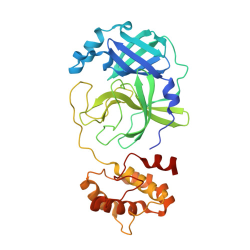
3C-like proteinase
A, B
309Severe acute respiratory syndrome coronavirus 2Mutation(s): 0 
Gene Names: rep1a-1b
EC: 3.4.22.69
UniProt
Find proteins for P0DTD1 (Severe acute respiratory syndrome coronavirus 2)
Explore P0DTD1 
Go to UniProtKB:  P0DTD1
Entity Groups  
Sequence Clusters30% Identity50% Identity70% Identity90% Identity95% Identity100% Identity
UniProt GroupP0DTD1
Sequence Annotations
Expand
  • Reference Sequence
Small Molecules
Ligands 3 Unique
IDChains Name / Formula / InChI Key2D Diagram3D Interactions
YKP
Query on YKP

Download Ideal Coordinates CCD File 
D [auth A],
E [auth B]
(1S,2S)-2-((S)-2-((((S)-1-(4,4-difluorocyclohexyl)ethoxy)carbonyl)amino)-4-methylpentanamido)-1-hydroxy-3-((S)-2-oxopyrrolidin-3-yl)propane-1-sulfonic acid
C22 H37 F2 N3 O8 S
YRNJEPAFOLVHMN-ZZKADIETSA-N
YKM
Query on YKM

Download Ideal Coordinates CCD File 
C [auth A],
F [auth B]
(1R,2S)-2-((S)-2-((((S)-1-(4,4-difluorocyclohexyl)ethoxy)carbonyl)amino)-4-methylpentanamido)-1-hydroxy-3-((S)-2-oxopyrrolidin-3-yl)propane-1-sulfonic acid
C22 H37 F2 N3 O8 S
YRNJEPAFOLVHMN-GINSCSNVSA-N
PG4
Query on PG4

Download Ideal Coordinates CCD File 
G [auth B]TETRAETHYLENE GLYCOL
C8 H18 O5
UWHCKJMYHZGTIT-UHFFFAOYSA-N
Experimental Data & Validation

Experimental Data

  • Method: X-RAY DIFFRACTION
  • Resolution: 1.60 Å
  • R-Value Free: 0.201 
  • R-Value Work: 0.178 
  • R-Value Observed: 0.179 
  • Space Group: P 1 21 1
Unit Cell:
Length ( Å )Angle ( ˚ )
a = 55.397α = 90
b = 98.967β = 108.34
c = 59.152γ = 90
Software Package:
Software NamePurpose
PHENIXrefinement
XDSdata reduction
Aimlessdata scaling
PHASERphasing
PDB_EXTRACTdata extraction

Structure Validation

View Full Validation Report


Ligand Structure Quality Assessment 


Entry History & Funding Information

Deposition Data


Funding OrganizationLocationGrant Number
National Institutes of Health/National Institute Of Allergy and Infectious Diseases (NIH/NIAID)United StatesR01AI109039

Revision History  (Full details and data files)

  • Version 1.0: 2021-03-24
    Type: Initial release
  • Version 1.1: 2021-12-15
    Changes: Database references
  • Version 1.2: 2022-01-05
    Changes: Database references
  • Version 1.3: 2023-10-18
    Changes: Data collection, Refinement description
  • Version 1.4: 2024-11-20
    Changes: Structure summary