7M3I

Structure of SARS-CoV-2 spike protein receptor binding domain in complex with a neutralizing antibody, CV2-75 Fab


Experimental Data Snapshot

  • Method: X-RAY DIFFRACTION
  • Resolution: 2.80 Å
  • R-Value Free: 
    0.281 (Depositor), 0.280 (DCC) 
  • R-Value Work: 
    0.245 (Depositor), 0.250 (DCC) 
  • R-Value Observed: 
    0.247 (Depositor) 

Starting Models: experimental
View more details

wwPDB Validation   3D Report Full Report


Ligand Structure Quality Assessment 

Created with Raphaël 2.3.0Worse 01 BetterLigand structure goodness of fit to experimental dataBest fitted NAGClick on this verticalbar to view details

This is version 1.3 of the entry. See complete history


Literature

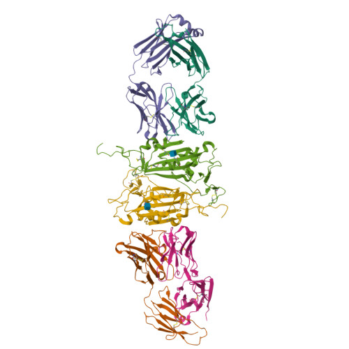
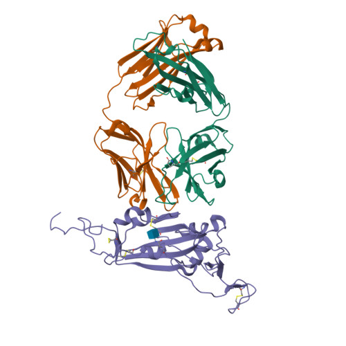
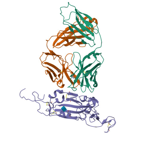
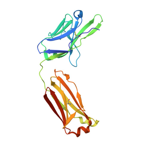
Isolation and characterization of cross-neutralizing coronavirus antibodies from COVID-19+ subjects.

Jennewein, M.F.MacCamy, A.J.Akins, N.R.Feng, J.Homad, L.J.Hurlburt, N.K.Seydoux, E.Wan, Y.H.Stuart, A.B.Edara, V.V.Floyd, K.Vanderheiden, A.Mascola, J.R.Doria-Rose, N.Wang, L.Yang, E.S.Chu, H.Y.Torres, J.L.Ozorowski, G.Ward, A.B.Whaley, R.E.Cohen, K.W.Pancera, M.McElrath, M.J.Englund, J.A.Finzi, A.Suthar, M.S.McGuire, A.T.Stamatatos, L.

(2021) Cell Rep 36: 109353-109353

  • DOI: https://doi.org/10.1016/j.celrep.2021.109353
  • Primary Citation of Related Structures:  
    7M3I

  • PubMed Abstract: 

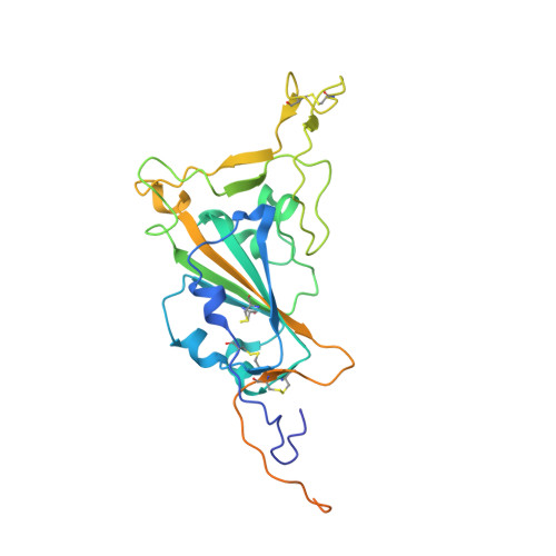
    SARS-CoV-2 is one of three coronaviruses that have crossed the animal-to-human barrier and caused widespread disease in the past two decades. The development of a universal human coronavirus vaccine could prevent future pandemics. We characterize 198 antibodies isolated from four COVID-19+ subjects and identify 14 SARS-CoV-2 neutralizing antibodies. One targets the N-terminal domain (NTD), one recognizes an epitope in S2, and 11 bind the receptor-binding domain (RBD). Three anti-RBD neutralizing antibodies cross-neutralize SARS-CoV-1 by effectively blocking binding of both the SARS-CoV-1 and SARS-CoV-2 RBDs to the ACE2 receptor. Using the K18-hACE transgenic mouse model, we demonstrate that the neutralization potency and antibody epitope specificity regulates the in vivo protective potential of anti-SARS-CoV-2 antibodies. All four cross-neutralizing antibodies neutralize the B.1.351 mutant strain. Thus, our study reveals that epitopes in S2 can serve as blueprints for the design of immunogens capable of eliciting cross-neutralizing coronavirus antibodies.


  • Organizational Affiliation

    Fred Hutchinson Cancer Research Center, Vaccines and Infectious Disease Division, Seattle, WA 98109, USA.


Macromolecules
Find similar proteins by:  (by identity cutoff)  |  3D Structure
Entity ID: 1
MoleculeChains Sequence LengthOrganismDetailsImage
CV2-75 Fab Heavy chainA,
D [auth H]
227Homo sapiensMutation(s): 0 
Entity Groups  
Sequence Clusters30% Identity50% Identity70% Identity90% Identity95% Identity100% Identity
Sequence Annotations
Expand
  • Reference Sequence
Find similar proteins by:  (by identity cutoff)  |  3D Structure
Entity ID: 2
MoleculeChains Sequence LengthOrganismDetailsImage
CV2-75 Fab Light chainB,
E [auth L]
214Homo sapiensMutation(s): 0 
Entity Groups  
Sequence Clusters30% Identity50% Identity70% Identity90% Identity95% Identity100% Identity
Sequence Annotations
Expand
  • Reference Sequence
Find similar proteins by:  (by identity cutoff)  |  3D Structure
Entity ID: 3
MoleculeChains Sequence LengthOrganismDetailsImage
Spike protein S1C,
F [auth R]
273Severe acute respiratory syndrome coronavirus 2Mutation(s): 0 
Gene Names: S2
UniProt
Find proteins for P0DTC2 (Severe acute respiratory syndrome coronavirus 2)
Explore P0DTC2 
Go to UniProtKB:  P0DTC2
Entity Groups  
Sequence Clusters30% Identity50% Identity70% Identity90% Identity95% Identity100% Identity
UniProt GroupP0DTC2
Glycosylation
Glycosylation Sites: 1Go to GlyGen: P0DTC2-1
Sequence Annotations
Expand
  • Reference Sequence
Small Molecules
Modified Residues  1 Unique
IDChains TypeFormula2D DiagramParent
PCA
Query on PCA
A,
D [auth H]
L-PEPTIDE LINKINGC5 H7 N O3GLN
Experimental Data & Validation

Experimental Data

  • Method: X-RAY DIFFRACTION
  • Resolution: 2.80 Å
  • R-Value Free:  0.281 (Depositor), 0.280 (DCC) 
  • R-Value Work:  0.245 (Depositor), 0.250 (DCC) 
  • R-Value Observed: 0.247 (Depositor) 
Space Group: P 21 21 21
Unit Cell:
Length ( Å )Angle ( ˚ )
a = 86.371α = 90
b = 127.519β = 90
c = 155.672γ = 90
Software Package:
Software NamePurpose
PHENIXrefinement
XDSdata reduction
Aimlessdata scaling
PHASERphasing

Structure Validation

View Full Validation Report


Ligand Structure Quality Assessment 

Created with Raphaël 2.3.0Worse 01 BetterLigand structure goodness of fit to experimental dataBest fitted NAGClick on this verticalbar to view details

Entry History 

Deposition Data

Revision History  (Full details and data files)

  • Version 1.0: 2021-05-12
    Type: Initial release
  • Version 1.1: 2021-07-28
    Changes: Database references
  • Version 1.2: 2023-10-18
    Changes: Data collection, Database references, Refinement description
  • Version 1.3: 2024-10-16
    Changes: Structure summary