7M6H

Structure of the SARS-CoV-2 S 2P trimer in complex with the human neutralizing antibody Fab fragment, BG7-20


Experimental Data Snapshot

  • Method: ELECTRON MICROSCOPY
  • Resolution: 4.00 Å
  • Aggregation State: PARTICLE 
  • Reconstruction Method: SINGLE PARTICLE 

wwPDB Validation   3D Report Full Report


This is version 1.2 of the entry. See complete history


Literature

B cell genomics behind cross-neutralization of SARS-CoV-2 variants and SARS-CoV.

Scheid, J.F.Barnes, C.O.Eraslan, B.Hudak, A.Keeffe, J.R.Cosimi, L.A.Brown, E.M.Muecksch, F.Weisblum, Y.Zhang, S.Delorey, T.Woolley, A.E.Ghantous, F.Park, S.M.Phillips, D.Tusi, B.Huey-Tubman, K.E.Cohen, A.A.Gnanapragasam, P.N.P.Rzasa, K.Hatziioanno, T.Durney, M.A.Gu, X.Tada, T.Landau, N.R.West Jr., A.P.Rozenblatt-Rosen, O.Seaman, M.S.Baden, L.R.Graham, D.B.Deguine, J.Bieniasz, P.D.Regev, A.Hung, D.Bjorkman, P.J.Xavier, R.J.

(2021) Cell 184: 3205

  • DOI: https://doi.org/10.1016/j.cell.2021.04.032
  • Primary Citation of Related Structures:  
    7M6D, 7M6E, 7M6F, 7M6G, 7M6H, 7M6I

  • PubMed Abstract: 

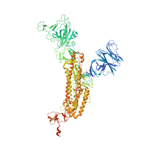
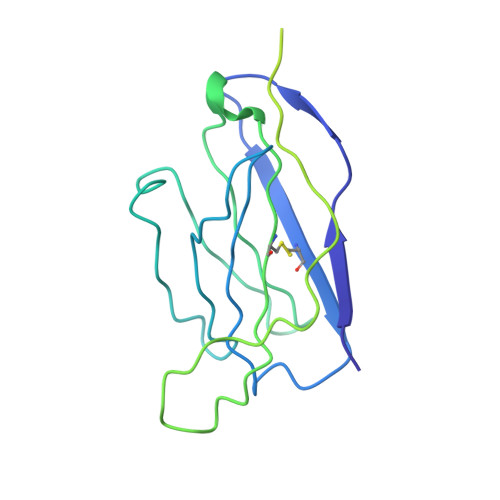
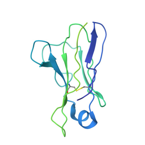
    Monoclonal antibodies (mAbs) are a focus in vaccine and therapeutic design to counteract severe acute respiratory syndrome coronavirus 2 (SARS-CoV-2) and its variants. Here, we combined B cell sorting with single-cell VDJ and RNA sequencing (RNA-seq) and mAb structures to characterize B cell responses against SARS-CoV-2. We show that the SARS-CoV-2-specific B cell repertoire consists of transcriptionally distinct B cell populations with cells producing potently neutralizing antibodies (nAbs) localized in two clusters that resemble memory and activated B cells. Cryo-electron microscopy structures of selected nAbs from these two clusters complexed with SARS-CoV-2 spike trimers show recognition of various receptor-binding domain (RBD) epitopes. One of these mAbs, BG10-19, locks the spike trimer in a closed conformation to potently neutralize SARS-CoV-2, the recently arising mutants B.1.1.7 and B.1.351, and SARS-CoV and cross-reacts with heterologous RBDs. Together, our results characterize transcriptional differences among SARS-CoV-2-specific B cells and uncover cross-neutralizing Ab targets that will inform immunogen and therapeutic design against coronaviruses.


  • Organizational Affiliation

    Broad Institute of the Massachusetts Institute of Technology and Harvard University, Cambridge, MA 02142, USA; Division of Gastroenterology, Massachusetts General Hospital and Harvard Medical School, Boston, MA 02114, USA; Center for Computational and Integrative Biology, Massachusetts General Hospital and Harvard Medical School, Boston, MA 02114, USA.


Macromolecules
Find similar proteins by:  (by identity cutoff)  |  3D Structure
Entity ID: 1
MoleculeChains Sequence LengthOrganismDetailsImage
Spike glycoprotein
A, B, C
1,259Severe acute respiratory syndrome coronavirus 2Mutation(s): 2 
Gene Names: S2
UniProt
Find proteins for P0DTC2 (Severe acute respiratory syndrome coronavirus 2)
Explore P0DTC2 
Go to UniProtKB:  P0DTC2
Entity Groups  
Sequence Clusters30% Identity50% Identity70% Identity90% Identity95% Identity100% Identity
UniProt GroupP0DTC2
Sequence Annotations
Expand
  • Reference Sequence
Find similar proteins by:  (by identity cutoff)  |  3D Structure
Entity ID: 2
MoleculeChains Sequence LengthOrganismDetailsImage
BG7-20 Fab Heavy Chain
D, E
233Homo sapiensMutation(s): 0 
Entity Groups  
Sequence Clusters30% Identity50% Identity70% Identity90% Identity95% Identity100% Identity
Sequence Annotations
Expand
  • Reference Sequence
Find similar proteins by:  (by identity cutoff)  |  3D Structure
Entity ID: 3
MoleculeChains Sequence LengthOrganismDetailsImage
BG7-20 Fab Light Chain
F, G
217Homo sapiensMutation(s): 0 
Entity Groups  
Sequence Clusters30% Identity50% Identity70% Identity90% Identity95% Identity100% Identity
Sequence Annotations
Expand
  • Reference Sequence
Small Molecules
Ligands 1 Unique
IDChains Name / Formula / InChI Key2D Diagram3D Interactions
NAG
Query on NAG

Download Ideal Coordinates CCD File 
AA [auth C]
BA [auth C]
CA [auth C]
DA [auth C]
EA [auth C]
AA [auth C],
BA [auth C],
CA [auth C],
DA [auth C],
EA [auth C],
FA [auth C],
GA [auth C],
H [auth A],
HA [auth C],
I [auth A],
IA [auth C],
J [auth A],
K [auth A],
L [auth A],
M [auth A],
N [auth A],
O [auth A],
P [auth A],
Q [auth A],
R [auth B],
S [auth B],
T [auth B],
U [auth B],
V [auth B],
W [auth B],
X [auth B],
Y [auth B],
Z [auth C]
2-acetamido-2-deoxy-beta-D-glucopyranose
C8 H15 N O6
OVRNDRQMDRJTHS-FMDGEEDCSA-N
Experimental Data & Validation

Experimental Data

  • Method: ELECTRON MICROSCOPY
  • Resolution: 4.00 Å
  • Aggregation State: PARTICLE 
  • Reconstruction Method: SINGLE PARTICLE 
EM Software:
TaskSoftware PackageVersion
MODEL REFINEMENTPHENIX1.19
RECONSTRUCTIONcryoSPARC2.14

Structure Validation

View Full Validation Report


Entry History & Funding Information

Deposition Data


Funding OrganizationLocationGrant Number
National Institutes of Health/National Institute Of Allergy and Infectious Diseases (NIH/NIAID)United StatesP01-AI138398-S1
National Institutes of Health/National Institute Of Allergy and Infectious Diseases (NIH/NIAID)United StatesP50 AI150464-13

Revision History  (Full details and data files)

  • Version 1.0: 2021-05-05
    Type: Initial release
  • Version 1.1: 2021-06-02
    Changes: Database references
  • Version 1.2: 2021-06-23
    Changes: Database references, Derived calculations