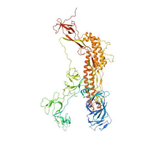
7MTD

Structure of aged SARS-CoV-2 S2P spike at pH 7.4


Experimental Data Snapshot

  • Method: ELECTRON MICROSCOPY
  • Resolution: 3.50 Å
  • Aggregation State: PARTICLE 
  • Reconstruction Method: SINGLE PARTICLE 

wwPDB Validation   3D Report Full Report


This is version 1.2 of the entry. See complete history


Literature

SARS-CoV-2 S2P spike ages through distinct states with altered immunogenicity.

Olia, A.S.Tsybovsky, Y.Chen, S.J.Liu, C.Nazzari, A.F.Ou, L.Wang, L.Kong, W.P.Leung, K.Liu, T.Stephens, T.Teng, I.T.Wang, S.Yang, E.S.Zhang, B.Zhang, Y.Zhou, T.Mascola, J.R.Kwong, P.D.

(2021) J Biol Chem 297: 101127-101127

  • DOI: https://doi.org/10.1016/j.jbc.2021.101127
  • Primary Citation of Related Structures:  
    7MTC, 7MTD, 7MTE

  • PubMed Abstract: 

    The SARS-CoV-2 spike is the primary target of virus-neutralizing antibodies and critical to the development of effective vaccines against COVID-19. Here, we demonstrate that the prefusion-stabilized two-proline "S2P" spike-widely employed for laboratory work and clinical studies-unfolds when stored at 4 °C, physiological pH, as observed by electron microscopy (EM) and differential scanning calorimetry, but that its trimeric, native-like conformation can be reacquired by low pH treatment. When stored for approximately 1 week, this unfolding does not significantly alter antigenic characteristics; however, longer storage diminishes antibody binding, and month-old spike elicits virtually no neutralization in mice despite inducing high ELISA-binding titers. Cryo-EM structures reveal the folded fraction of spike to decrease with aging; however, its structure remains largely similar, although with varying mobility of the receptor-binding domain. Thus, the SARS-CoV-2 spike is susceptible to unfolding, which affects immunogenicity, highlighting the need to monitor its integrity.


  • Organizational Affiliation

    Vaccine Research Center, National Institute of Allergy and Infectious Diseases, National Institutes of Health, Bethesda, Maryland, USA.


Macromolecules
Find similar proteins by:  (by identity cutoff)  |  3D Structure
Entity ID: 1
MoleculeChains Sequence LengthOrganismDetailsImage
Spike glycoprotein
A, B, C
1,281Severe acute respiratory syndrome coronavirus 2Mutation(s): 0 
Gene Names: S2
UniProt
Find proteins for P0DTC2 (Severe acute respiratory syndrome coronavirus 2)
Explore P0DTC2 
Go to UniProtKB:  P0DTC2
Entity Groups  
Sequence Clusters30% Identity50% Identity70% Identity90% Identity95% Identity100% Identity
UniProt GroupP0DTC2
Glycosylation
Glycosylation Sites: 9Go to GlyGen: P0DTC2-1
Sequence Annotations
Expand
  • Reference Sequence
Oligosaccharides

Help

Entity ID: 2
MoleculeChains Length2D Diagram Glycosylation3D Interactions
2-acetamido-2-deoxy-beta-D-glucopyranose-(1-4)-2-acetamido-2-deoxy-beta-D-glucopyranose
D, E, F, G, H
D, E, F, G, H, I, J, K, L, M, N, O, P, Q
2N-Glycosylation
Glycosylation Resources
GlyTouCan:  G42666HT
GlyCosmos:  G42666HT
GlyGen:  G42666HT
Small Molecules
Ligands 1 Unique
IDChains Name / Formula / InChI Key2D Diagram3D Interactions
NAG
Query on NAG

Download Ideal Coordinates CCD File 
AA [auth C]
BA [auth C]
CA [auth C]
DA [auth C]
R [auth A]
AA [auth C],
BA [auth C],
CA [auth C],
DA [auth C],
R [auth A],
S [auth A],
T [auth A],
U [auth A],
V [auth B],
W [auth B],
X [auth B],
Y [auth B],
Z [auth B]
2-acetamido-2-deoxy-beta-D-glucopyranose
C8 H15 N O6
OVRNDRQMDRJTHS-FMDGEEDCSA-N
Experimental Data & Validation

Experimental Data

  • Method: ELECTRON MICROSCOPY
  • Resolution: 3.50 Å
  • Aggregation State: PARTICLE 
  • Reconstruction Method: SINGLE PARTICLE 
EM Software:
TaskSoftware PackageVersion
RECONSTRUCTIONRELION3.1

Structure Validation

View Full Validation Report


Entry History 

Deposition Data

Revision History  (Full details and data files)

  • Version 1.0: 2021-09-15
    Type: Initial release
  • Version 1.1: 2021-10-13
    Changes: Database references
  • Version 1.2: 2024-10-23
    Changes: Data collection, Structure summary