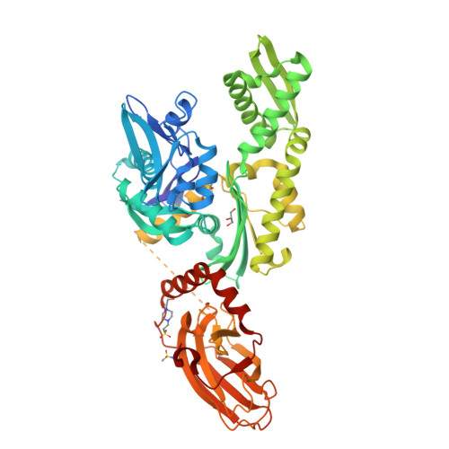
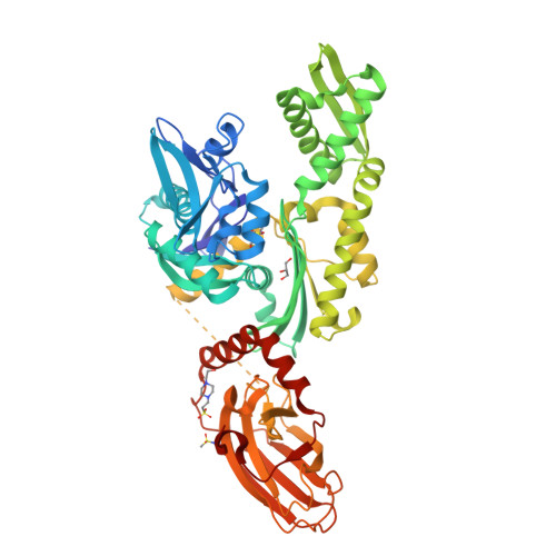
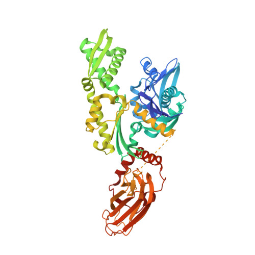
7ODI | pdb_00007odi

Crystal structure of bovine Hsc70(aa1-554)E213A/D214A in complex with methanesulfonamide


Experimental Data Snapshot

  • Method: X-RAY DIFFRACTION
  • Resolution: 1.83 Å
  • R-Value Free: 
    0.245 (Depositor), 0.250 (DCC) 
  • R-Value Work: 
    0.230 (Depositor), 0.230 (DCC) 
  • R-Value Observed: 
    0.231 (Depositor) 

Starting Model: experimental
View more details

wwPDB Validation   3D Report Full Report


Ligand Structure Quality Assessment 

Created with Raphaël 2.3.0Worse 01 BetterLigand structure goodness of fit to experimental dataBest fitted V7ZClick on this verticalbar to view details

This is version 1.2 of the entry. See complete history


Literature

Combined In-Solution Fragment Screening and Crystallographic Binding-Mode Analysis with a Two-Domain Hsp70 Construct.

Zehe, M.Kehrein, J.Schollmayer, C.Plank, C.Kovacs, H.Merino Asumendi, E.Holzgrabe, U.Grimm, C.Sotriffer, C.

(2024) ACS Chem Biol 

  • DOI: https://doi.org/10.1021/acschembio.3c00589
  • Primary Citation of Related Structures:  
    6H54, 7O6R, 7ODB, 7ODD, 7ODI, 7PLK

  • PubMed Abstract: 

    Heat shock protein 70 (Hsp70) isoforms are key players in the regulation of protein homeostasis and cell death pathways and are therefore attractive targets in cancer research. Developing nucleotide-competitive inhibitors or allosteric modulators, however, has turned out to be very challenging for this protein family, and no Hsp70-directed therapeutics have so far become available. As the field could profit from alternative starting points for inhibitor development, we present the results of a fragment-based screening approach on a two-domain Hsp70 construct using in-solution NMR methods, together with X-ray-crystallographic investigations and mixed-solvent molecular dynamics simulations. The screening protocol resulted in hits on both domains. In particular, fragment binding in a deeply buried pocket at the substrate-binding domain could be detected. The corresponding site is known to be important for communication between the nucleotide-binding and substrate-binding domains of Hsp70 proteins. The main fragment identified at this position also offers an interesting starting point for the development of a dual Hsp70/Hsp90 inhibitor.


  • Organizational Affiliation

    University of Würzburg, Institute of Pharmacy and Food Chemistry, Am Hubland, DE-97074 Würzburg, Germany.


Macromolecules
Find similar proteins by:  (by identity cutoff)  |  3D Structure
Entity ID: 1
MoleculeChains Sequence LengthOrganismDetailsImage
Heat shock cognate 71 kDa protein554Bos taurusMutation(s): 2 
Gene Names: HSPA8HSC70
EC: 3.6.4.10
UniProt
Find proteins for P19120 (Bos taurus)
Explore P19120 
Go to UniProtKB:  P19120
Entity Groups  
Sequence Clusters30% Identity50% Identity70% Identity90% Identity95% Identity100% Identity
UniProt GroupP19120
Sequence Annotations
Expand
  • Reference Sequence
Small Molecules
Ligands 4 Unique
IDChains Name / Formula / InChI Key2D Diagram3D Interactions
EPE
Query on EPE

Download Ideal Coordinates CCD File 
B [auth A]4-(2-HYDROXYETHYL)-1-PIPERAZINE ETHANESULFONIC ACID
C8 H18 N2 O4 S
JKMHFZQWWAIEOD-UHFFFAOYSA-N
V7Z (Subject of Investigation/LOI)
Query on V7Z

Download Ideal Coordinates CCD File 
C [auth A],
D [auth A]
methanesulfonamide
C H5 N O2 S
HNQIVZYLYMDVSB-UHFFFAOYSA-N
GOL
Query on GOL

Download Ideal Coordinates CCD File 
E [auth A],
F [auth A]
GLYCEROL
C3 H8 O3
PEDCQBHIVMGVHV-UHFFFAOYSA-N
NA
Query on NA

Download Ideal Coordinates CCD File 
G [auth A]SODIUM ION
Na
FKNQFGJONOIPTF-UHFFFAOYSA-N
Experimental Data & Validation

Experimental Data

  • Method: X-RAY DIFFRACTION
  • Resolution: 1.83 Å
  • R-Value Free:  0.245 (Depositor), 0.250 (DCC) 
  • R-Value Work:  0.230 (Depositor), 0.230 (DCC) 
  • R-Value Observed: 0.231 (Depositor) 
Space Group: P 1 21 1
Unit Cell:
Length ( Å )Angle ( ˚ )
a = 65.847α = 90
b = 49.566β = 99.837
c = 86.716γ = 90
Software Package:
Software NamePurpose
PHENIXrefinement
MxCuBEdata collection
Cootmodel building

Structure Validation

View Full Validation Report


Ligand Structure Quality Assessment 

Created with Raphaël 2.3.0Worse 01 BetterLigand structure goodness of fit to experimental dataBest fitted V7ZClick on this verticalbar to view details

Entry History 

Deposition Data

Revision History  (Full details and data files)

  • Version 1.0: 2022-06-01
    Type: Initial release
  • Version 1.1: 2024-01-31
    Changes: Data collection, Refinement description
  • Version 1.2: 2024-02-14
    Changes: Database references