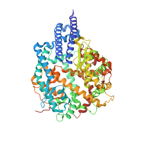
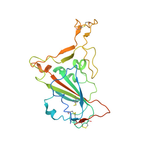
Evidence of SARS-CoV-2 Direct Evolution in R. affinis Bats Driven by Affinity and Dynamics Optimization of the Spike Protein
Castelli, M., Scietti, L., Faravelli, S., Clementi, N., Forneris, F., Mancini, N.To be published.
Experimental Data Snapshot
Starting Model: experimental
View more details
Entity ID: 1 | |||||
|---|---|---|---|---|---|
| Molecule | Chains | Sequence Length | Organism | Details | Image |
| Processed angiotensin-converting enzyme 2 | 602 | Homo sapiens | Mutation(s): 0 Gene Names: ACE2, UNQ868/PRO1885 EC: 3.4.17 (UniProt), 3.4.17.23 (UniProt) |  | |
UniProt & NIH Common Fund Data Resources | |||||
Find proteins for Q9BYF1 (Homo sapiens) Explore Q9BYF1 Go to UniProtKB: Q9BYF1 | |||||
PHAROS: Q9BYF1 GTEx: ENSG00000130234 | |||||
Entity Groups | |||||
| Sequence Clusters | 30% Identity50% Identity70% Identity90% Identity95% Identity100% Identity | ||||
| UniProt Group | Q9BYF1 | ||||
Glycosylation | |||||
| Glycosylation Sites: 4 | Go to GlyGen: Q9BYF1-1 | ||||
Sequence AnnotationsExpand | |||||
| |||||
Entity ID: 2 | |||||
|---|---|---|---|---|---|
| Molecule | Chains | Sequence Length | Organism | Details | Image |
| Spike glycoprotein | 233 | Bat coronavirus RaTG13 | Mutation(s): 0 |  | |
Entity Groups | |||||
| Sequence Clusters | 30% Identity50% Identity70% Identity90% Identity95% Identity100% Identity | ||||
Glycosylation | |||||
| Glycosylation Sites: 1 | |||||
Sequence AnnotationsExpand | |||||
| |||||
| Ligands 1 Unique | |||||
|---|---|---|---|---|---|
| ID | Chains | Name / Formula / InChI Key | 2D Diagram | 3D Interactions | |
| NAG Query on NAG | M [auth C], N [auth D], O [auth D] | 2-acetamido-2-deoxy-beta-D-glucopyranose C8 H15 N O6 OVRNDRQMDRJTHS-FMDGEEDCSA-N |  | ||
| Length ( Å ) | Angle ( ˚ ) |
|---|---|
| a = 83.557 | α = 90 |
| b = 131.214 | β = 99.77 |
| c = 115.725 | γ = 90 |
| Software Name | Purpose |
|---|---|
| PHENIX | refinement |
| PDB_EXTRACT | data extraction |
| xia2 | data reduction |
| Aimless | data scaling |
| PHASER | phasing |
| Funding Organization | Location | Grant Number |
|---|---|---|
| The Giovanni Armenise-Harvard Foundation | United States | CDA2013 |
| Italian Association for Cancer Research | Italy | MFAG 20075 |
| Mizutani Foundation for Glycoscience | Japan | 200039 |
| NATO Science for Peace and Security Program | Belgium | SPS.MYP G5701 |
| Velux Stiftung | Switzerland | 1375 |
| Italian Ministry of Education | Italy | PRIN 2017 2017RPHBCW_001 |
| Italian Ministry of Education | Italy | Department of Excellence 2018-2022 |