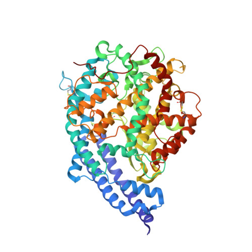
7PKI

Crystal structure of human ACE2 bound to the spike receptor-binding domain from a cave bat sarbecovirus closely related to SARS-CoV-2.


Experimental Data Snapshot

  • Method: X-RAY DIFFRACTION
  • Resolution: 2.94 Å
  • R-Value Free: 0.227 
  • R-Value Work: 0.214 
  • R-Value Observed: 0.215 

wwPDB Validation   3D Report Full Report


Ligand Structure Quality Assessment 


This is version 1.2 of the entry. See complete history


Literature

Bat coronaviruses related to SARS-CoV-2 and infectious for human cells.

Temmam, S.Vongphayloth, K.Baquero, E.Munier, S.Bonomi, M.Regnault, B.Douangboubpha, B.Karami, Y.Chretien, D.Sanamxay, D.Xayaphet, V.Paphaphanh, P.Lacoste, V.Somlor, S.Lakeomany, K.Phommavanh, N.Perot, P.Dehan, O.Amara, F.Donati, F.Bigot, T.Nilges, M.Rey, F.A.van der Werf, S.Brey, P.T.Eloit, M.

(2022) Nature 604: 330-336

  • DOI: https://doi.org/10.1038/s41586-022-04532-4
  • Primary Citation of Related Structures:  
    7PKI

  • PubMed Abstract: 

    The animal reservoir of SARS-CoV-2 is unknown despite reports of SARS-CoV-2-related viruses in Asian Rhinolophus bats 1-4 , including the closest virus from R. affinis, RaTG13 (refs. 5,6 ), and pangolins 7-9 . SARS-CoV-2 has a mosaic genome, to which different progenitors contribute. The spike sequence determines the binding affinity and accessibility of its receptor-binding domain to the cellular angiotensin-converting enzyme 2 (ACE2) receptor and is responsible for host range 10-12 . SARS-CoV-2 progenitor bat viruses genetically close to SARS-CoV-2 and able to enter human cells through a human ACE2 (hACE2) pathway have not yet been identified, although they would be key in understanding the origin of the epidemic. Here we show that such viruses circulate in cave bats living in the limestone karstic terrain in northern Laos, in the Indochinese peninsula. We found that the receptor-binding domains of these viruses differ from that of SARS-CoV-2 by only one or two residues at the interface with ACE2, bind more efficiently to the hACE2 protein than that of the SARS-CoV-2 strain isolated in Wuhan from early human cases, and mediate hACE2-dependent entry and replication in human cells, which is inhibited by antibodies that neutralize SARS-CoV-2. None of these bat viruses contains a furin cleavage site in the spike protein. Our findings therefore indicate that bat-borne SARS-CoV-2-like viruses that are potentially infectious for humans circulate in Rhinolophus spp. in the Indochinese peninsula.


  • Organizational Affiliation

    Institut Pasteur, Université de Paris, Pathogen Discovery Laboratory, Paris, France.


Macromolecules
Find similar proteins by:  (by identity cutoff)  |  3D Structure
Entity ID: 1
MoleculeChains Sequence LengthOrganismDetailsImage
Processed angiotensin-converting enzyme 2596Homo sapiensMutation(s): 0 
Gene Names: ACE2UNQ868/PRO1885
UniProt & NIH Common Fund Data Resources
Find proteins for Q9BYF1 (Homo sapiens)
Explore Q9BYF1 
Go to UniProtKB:  Q9BYF1
PHAROS:  Q9BYF1
GTEx:  ENSG00000130234 
Entity Groups  
Sequence Clusters30% Identity50% Identity70% Identity90% Identity95% Identity100% Identity
UniProt GroupQ9BYF1
Sequence Annotations
Expand
  • Reference Sequence
Find similar proteins by:  (by identity cutoff)  |  3D Structure
Entity ID: 2
MoleculeChains Sequence LengthOrganismDetailsImage
BANAL 236 coronavirus spike receptor-binding domainB [auth E]197SarbecovirusMutation(s): 0 
Gene Names: Spike
UniProt
Find proteins for P0DTC2 (Severe acute respiratory syndrome coronavirus 2)
Explore P0DTC2 
Go to UniProtKB:  P0DTC2
Entity Groups  
Sequence Clusters30% Identity50% Identity70% Identity90% Identity95% Identity100% Identity
UniProt GroupP0DTC2
Sequence Annotations
Expand
  • Reference Sequence
Oligosaccharides

Help

Entity ID: 3
MoleculeChains Length2D Diagram Glycosylation3D Interactions
2-acetamido-2-deoxy-beta-D-glucopyranose-(1-4)-2-acetamido-2-deoxy-beta-D-glucopyranoseC [auth B]2N-Glycosylation
Glycosylation Resources
GlyTouCan:  G42666HT
GlyCosmos:  G42666HT
GlyGen:  G42666HT
Small Molecules
Ligands 7 Unique
IDChains Name / Formula / InChI Key2D Diagram3D Interactions
NAG
Query on NAG

Download Ideal Coordinates CCD File 
D [auth A]
E [auth A]
F [auth A]
G [auth A]
O [auth E]
D [auth A],
E [auth A],
F [auth A],
G [auth A],
O [auth E],
P [auth E]
2-acetamido-2-deoxy-beta-D-glucopyranose
C8 H15 N O6
OVRNDRQMDRJTHS-FMDGEEDCSA-N
TRS
Query on TRS

Download Ideal Coordinates CCD File 
K [auth A]2-AMINO-2-HYDROXYMETHYL-PROPANE-1,3-DIOL
C4 H12 N O3
LENZDBCJOHFCAS-UHFFFAOYSA-O
PEG
Query on PEG

Download Ideal Coordinates CCD File 
L [auth A],
M [auth A],
T [auth E],
U [auth E],
V [auth E]
DI(HYDROXYETHYL)ETHER
C4 H10 O3
MTHSVFCYNBDYFN-UHFFFAOYSA-N
SO4
Query on SO4

Download Ideal Coordinates CCD File 
H [auth A],
I [auth A],
J [auth A],
Q [auth E]
SULFATE ION
O4 S
QAOWNCQODCNURD-UHFFFAOYSA-L
GOL
Query on GOL

Download Ideal Coordinates CCD File 
W [auth E]GLYCEROL
C3 H8 O3
PEDCQBHIVMGVHV-UHFFFAOYSA-N
ZN
Query on ZN

Download Ideal Coordinates CCD File 
N [auth A]ZINC ION
Zn
PTFCDOFLOPIGGS-UHFFFAOYSA-N
CL
Query on CL

Download Ideal Coordinates CCD File 
R [auth E],
S [auth E]
CHLORIDE ION
Cl
VEXZGXHMUGYJMC-UHFFFAOYSA-M
Experimental Data & Validation

Experimental Data

  • Method: X-RAY DIFFRACTION
  • Resolution: 2.94 Å
  • R-Value Free: 0.227 
  • R-Value Work: 0.214 
  • R-Value Observed: 0.215 
  • Space Group: P 65 2 2
Unit Cell:
Length ( Å )Angle ( ˚ )
a = 341.535α = 90
b = 341.535β = 90
c = 68.153γ = 120
Software Package:
Software NamePurpose
PHENIXrefinement
XDSdata reduction
XDSdata scaling
autoBUSTERphasing

Structure Validation

View Full Validation Report


Ligand Structure Quality Assessment 


Entry History & Funding Information

Deposition Data


Funding OrganizationLocationGrant Number
Pasteur InstituteFranceAlloSpike

Revision History  (Full details and data files)

  • Version 1.0: 2022-01-19
    Type: Initial release
  • Version 1.1: 2023-02-01
    Changes: Database references
  • Version 1.2: 2024-02-07
    Changes: Data collection, Refinement description