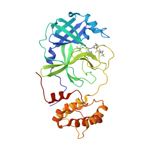
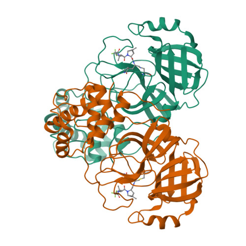
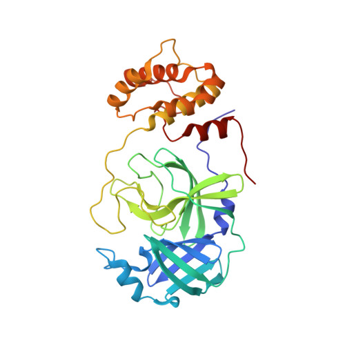
7TE0

Structure of the SARS-CoV-2 main protease in complex with inhibitor PF-07321332


Experimental Data Snapshot

  • Method: X-RAY DIFFRACTION
  • Resolution: 2.00 Å
  • R-Value Free: 0.248 
  • R-Value Work: 0.199 
  • R-Value Observed: 0.202 

Starting Model: experimental
View more details

wwPDB Validation   3D Report Full Report


Ligand Structure Quality Assessment 


This is version 1.3 of the entry. See complete history


Literature

Evolutionary and Structural Insights about Potential SARS-CoV-2 Evasion of Nirmatrelvir.

Yang, K.S.Leeuwon, S.Z.Xu, S.Liu, W.R.

(2022) J Med Chem 65: 8686-8698

  • DOI: https://doi.org/10.1021/acs.jmedchem.2c00404
  • Primary Citation of Related Structures:  
    7TE0

  • PubMed Abstract: 

    The U.S. FDA approval of PAXLOVID, a combination therapy of nirmatrelvir and ritonavir has significantly boosted our morale in fighting the COVID-19 pandemic. Nirmatrelvir is an inhibitor of the main protease (M Pro ) of SARS-CoV-2. Since many SARS-CoV-2 variants that resist vaccines and antibodies have emerged, a concern of acquired viral resistance to nirmatrelvir naturally arises. Here, possible mutations in M Pro to confer viral evasion of nirmatrelvir are analyzed and discussed from both evolutionary and structural standpoints. The analysis indicates that those mutations will likely reside in the whole aa45-51 helical region and residues including M165, L167, P168, R188, and Q189. Relevant mutations have also been observed in existing SARS-CoV-2 samples. Implications of this analysis to the fight against future drug-resistant viral variants and the development of broad-spectrum antivirals are discussed as well.


  • Organizational Affiliation

    Texas A&M Drug Discovery Laboratory, Department of Chemistry, Texas A&M University, College Station, Texas 77843, United States.


Macromolecules
Find similar proteins by:  (by identity cutoff)  |  3D Structure
Entity ID: 1
MoleculeChains Sequence LengthOrganismDetailsImage
3C-like proteinase306Severe acute respiratory syndrome coronavirus 2Mutation(s): 0 
Gene Names: rep1a-1b
EC: 3.4.22.69
UniProt
Find proteins for P0DTD1 (Severe acute respiratory syndrome coronavirus 2)
Explore P0DTD1 
Go to UniProtKB:  P0DTD1
Entity Groups  
Sequence Clusters30% Identity50% Identity70% Identity90% Identity95% Identity100% Identity
UniProt GroupP0DTD1
Sequence Annotations
Expand
  • Reference Sequence
Small Molecules
Ligands 1 Unique
IDChains Name / Formula / InChI Key2D Diagram3D Interactions
4WI (Subject of Investigation/LOI)
Query on 4WI

Download Ideal Coordinates CCD File 
B [auth A](1R,2S,5S)-N-{(1E,2S)-1-imino-3-[(3S)-2-oxopyrrolidin-3-yl]propan-2-yl}-6,6-dimethyl-3-[3-methyl-N-(trifluoroacetyl)-L-valyl]-3-azabicyclo[3.1.0]hexane-2-carboxamide
C23 H34 F3 N5 O4
WDVIRQQKRMIXGS-XIFHJVQQSA-N
Experimental Data & Validation

Experimental Data

  • Method: X-RAY DIFFRACTION
  • Resolution: 2.00 Å
  • R-Value Free: 0.248 
  • R-Value Work: 0.199 
  • R-Value Observed: 0.202 
  • Space Group: I 1 2 1
Unit Cell:
Length ( Å )Angle ( ˚ )
a = 51.637α = 90
b = 81.351β = 96.642
c = 89.746γ = 90
Software Package:
Software NamePurpose
PHENIXrefinement
PROTEUM PLUSdata reduction
Aimlessdata scaling
PHENIXphasing

Structure Validation

View Full Validation Report


Ligand Structure Quality Assessment 


Entry History & Funding Information

Deposition Data


Funding OrganizationLocationGrant Number
Welch FoundationUnited States--

Revision History  (Full details and data files)

  • Version 1.0: 2022-01-12
    Type: Initial release
  • Version 1.1: 2022-07-27
    Changes: Database references
  • Version 1.2: 2023-10-18
    Changes: Data collection, Refinement description
  • Version 1.3: 2024-10-30
    Changes: Structure summary