7TGR

Structure of SARS-CoV-2 main protease in complex with GC376


Experimental Data Snapshot

  • Method: X-RAY DIFFRACTION
  • Resolution: 1.68 Å
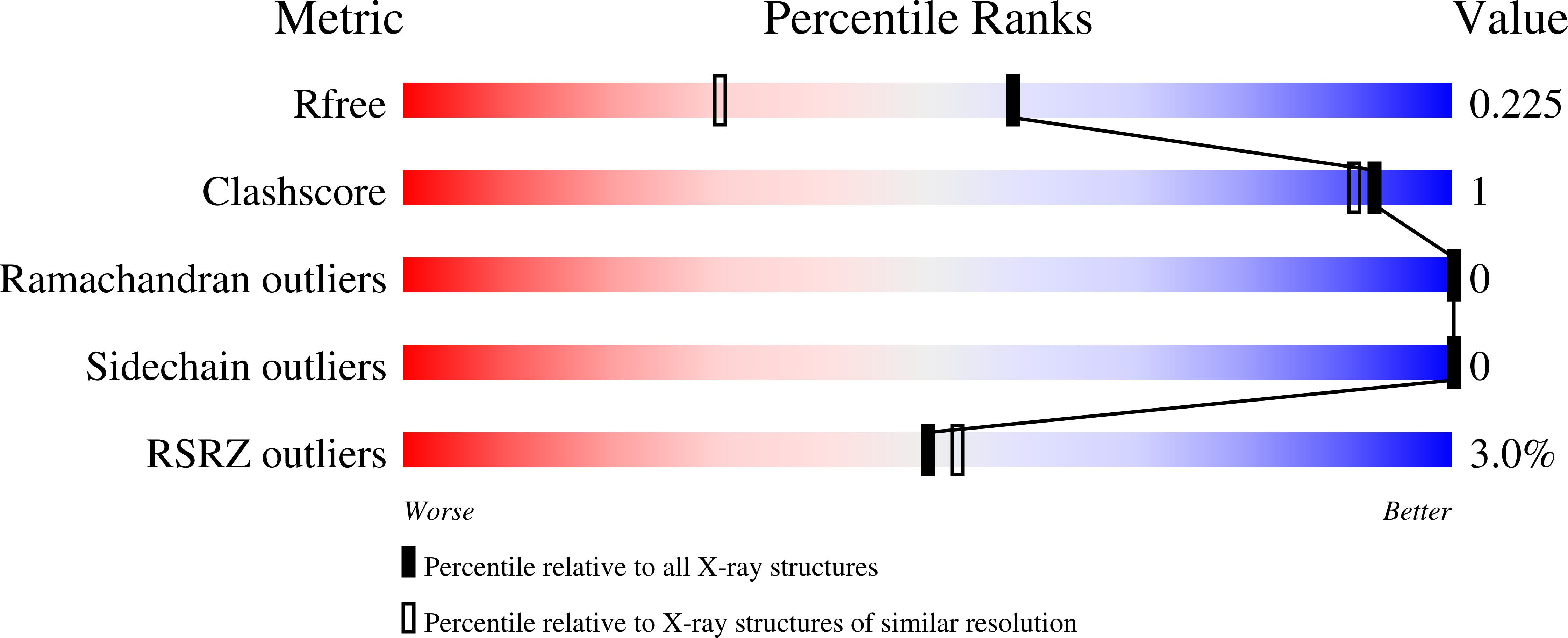
  • R-Value Free: 0.225 
  • R-Value Work: 0.182 
  • R-Value Observed: 0.184 

Starting Model: experimental
View more details

wwPDB Validation   3D Report Full Report


Ligand Structure Quality Assessment 


This is version 1.4 of the entry. See complete history


Literature

Gain-of-Signal Assays for Probing Inhibition of SARS-CoV-2 M pro /3CL pro in Living Cells.

Moghadasi, S.A.Esler, M.A.Otsuka, Y.Becker, J.T.Moraes, S.N.Anderson, C.B.Chamakuri, S.Belica, C.Wick, C.Harki, D.A.Young, D.W.Scampavia, L.Spicer, T.P.Shi, K.Aihara, H.Brown, W.L.Harris, R.S.

(2022) mBio 13: e0078422-e0078422

  • DOI: https://doi.org/10.1128/mbio.00784-22
  • Primary Citation of Related Structures:  
    7TGR

  • PubMed Abstract: 

    The main protease, M pro , of SARS-CoV-2 is required to cleave the viral polyprotein into precise functional units for virus replication and pathogenesis. Here, we report quantitative reporters for M pro function in living cells in which protease inhibition by genetic or chemical methods results in robust signal readouts by fluorescence (enhanced green fluorescent protein [eGFP]) or bioluminescence (firefly luciferase). These gain-of-signal systems are scalable to high-throughput platforms for quantitative discrimination between M pro mutants and/or inhibitor potencies as evidenced by validation of several reported inhibitors. Additional utility is shown by single M pro amino acid variants and structural information combining to demonstrate that both inhibitor conformational dynamics and amino acid differences are able to influence inhibitor potency. We further show that a recent variant of concern (Omicron) has an unchanged response to a clinically approved drug, nirmatrelvir, whereas proteases from divergent coronavirus species show differential susceptibility. Together, we demonstrate that these gain-of-signal systems serve as robust, facile, and scalable assays for live cell quantification of M pro inhibition, which will help expedite the development of next-generation antivirals and enable the rapid testing of emerging variants. IMPORTANCE The main protease, M pro , of SARS-CoV-2 is an essential viral protein required for the earliest steps of infection. It is therefore an attractive target for antiviral drug development. Here, we report the development and implementation of two complementary cell-based systems for quantification of M pro inhibition by genetic or chemical approaches. The first is fluorescence based (eGFP), and the second is luminescence based (firefly luciferase). Importantly, both systems rely upon gain-of-signal readouts such that stronger inhibitors yield higher fluorescent or luminescent signal. The high versatility and utility of these systems are demonstrated by characterizing M pro mutants and natural variants, including Omicron, as well as a panel of existing inhibitors. These systems rapidly, safely, and sensitively identify M pro variants with altered susceptibilities to inhibition, triage-nonspecific, or off-target molecules and validate bona fide inhibitors, with the most potent thus far being the first-in-class drug nirmatrelvir.


  • Organizational Affiliation

    Department of Biochemistry, Molecular Biology and Biophysics, University of Minnesotagrid.17635.36, Minneapolis, Minnesota, USA.


Macromolecules
Find similar proteins by:  (by identity cutoff)  |  3D Structure
Entity ID: 1
MoleculeChains Sequence LengthOrganismDetailsImage
3C-like proteinase304Severe acute respiratory syndrome coronavirus 2Mutation(s): 0 
Gene Names: rep1a-1b
EC: 3.4.22.69
UniProt
Find proteins for P0DTD1 (Severe acute respiratory syndrome coronavirus 2)
Explore P0DTD1 
Go to UniProtKB:  P0DTD1
Entity Groups  
Sequence Clusters30% Identity50% Identity70% Identity90% Identity95% Identity100% Identity
UniProt GroupP0DTD1
Sequence Annotations
Expand
  • Reference Sequence
Small Molecules
Ligands 6 Unique
IDChains Name / Formula / InChI Key2D Diagram3D Interactions
B1S (Subject of Investigation/LOI)
Query on B1S

Download Ideal Coordinates CCD File 
C [auth A](1R,2S)-2-({N-[(benzyloxy)carbonyl]-L-leucyl}amino)-1-hydroxy-3-[(3S)-2-oxopyrrolidin-3-yl]propane-1-sulfonic acid
C21 H31 N3 O8 S
BSPZFJDYQHDZNR-OGNFBWPZSA-N
K36 (Subject of Investigation/LOI)
Query on K36

Download Ideal Coordinates CCD File 
B [auth A](1S,2S)-2-({N-[(benzyloxy)carbonyl]-L-leucyl}amino)-1-hydroxy-3-[(3S)-2-oxopyrrolidin-3-yl]propane-1-sulfonic acid
C21 H31 N3 O8 S
BSPZFJDYQHDZNR-HTCLRFROSA-N
EDO
Query on EDO

Download Ideal Coordinates CCD File 
D [auth A]1,2-ETHANEDIOL
C2 H6 O2
LYCAIKOWRPUZTN-UHFFFAOYSA-N
K
Query on K

Download Ideal Coordinates CCD File 
G [auth A]POTASSIUM ION
K
NPYPAHLBTDXSSS-UHFFFAOYSA-N
CL
Query on CL

Download Ideal Coordinates CCD File 
E [auth A],
F [auth A]
CHLORIDE ION
Cl
VEXZGXHMUGYJMC-UHFFFAOYSA-M
NA
Query on NA

Download Ideal Coordinates CCD File 
H [auth A]SODIUM ION
Na
FKNQFGJONOIPTF-UHFFFAOYSA-N
Biologically Interesting Molecules (External Reference) 1 Unique
Experimental Data & Validation

Experimental Data

  • Method: X-RAY DIFFRACTION
  • Resolution: 1.68 Å
  • R-Value Free: 0.225 
  • R-Value Work: 0.182 
  • R-Value Observed: 0.184 
  • Space Group: C 1 2 1
Unit Cell:
Length ( Å )Angle ( ˚ )
a = 114.565α = 90
b = 52.939β = 102.55
c = 45.904γ = 90
Software Package:
Software NamePurpose
PHENIXrefinement
XDSdata reduction
Aimlessdata scaling
PHENIXphasing

Structure Validation

View Full Validation Report


Ligand Structure Quality Assessment 


Entry History & Funding Information

Deposition Data


Funding OrganizationLocationGrant Number
National Institutes of Health/National Institute Of Allergy and Infectious Diseases (NIH/NIAID)United StatesR37-AI064064
National Institutes of Health/National Cancer Institute (NIH/NCI)United StatesP01-CA214279

Revision History  (Full details and data files)

  • Version 1.0: 2022-05-04
    Type: Initial release
  • Version 1.1: 2022-07-13
    Changes: Database references
  • Version 1.2: 2022-07-20
    Changes: Database references
  • Version 1.3: 2023-10-18
    Changes: Data collection, Refinement description
  • Version 1.4: 2024-11-20
    Changes: Structure summary