7US6

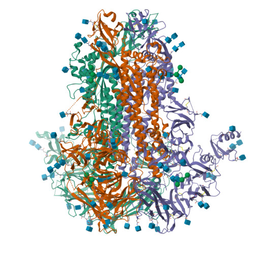
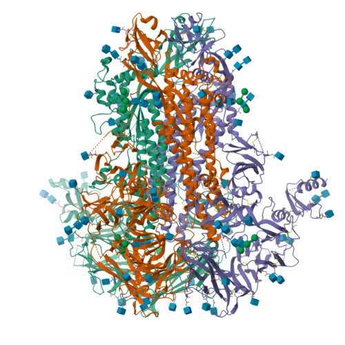
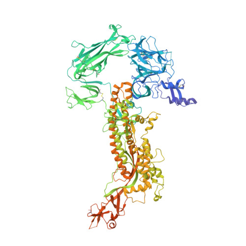
Structure of the human coronavirus CCoV-HuPn-2018 spike glycoprotein with domain 0 in the proximal conformation


Experimental Data Snapshot

  • Method: ELECTRON MICROSCOPY
  • Resolution: 3.80 Å
  • Aggregation State: PARTICLE 
  • Reconstruction Method: SINGLE PARTICLE 

wwPDB Validation   3D Report Full Report


This is version 1.0 of the entry. See complete history


Literature

Structure, receptor recognition, and antigenicity of the human coronavirus CCoV-HuPn-2018 spike glycoprotein.

Tortorici, M.A.Walls, A.C.Joshi, A.Park, Y.J.Eguia, R.T.Miranda, M.C.Kepl, E.Dosey, A.Stevens-Ayers, T.Boeckh, M.J.Telenti, A.Lanzavecchia, A.King, N.P.Corti, D.Bloom, J.D.Veesler, D.

(2022) Cell 185: 2279-2291.e17

  • DOI: https://doi.org/10.1016/j.cell.2022.05.019
  • Primary Citation of Related Structures:  
    7U0L, 7US6, 7US9, 7USA, 7USB

  • PubMed Abstract: 

    The isolation of CCoV-HuPn-2018 from a child respiratory swab indicates that more coronaviruses are spilling over to humans than previously appreciated. We determined the structures of the CCoV-HuPn-2018 spike glycoprotein trimer in two distinct conformational states and showed that its domain 0 recognizes sialosides. We identified that the CCoV-HuPn-2018 spike binds canine, feline, and porcine aminopeptidase N (APN) orthologs, which serve as entry receptors, and determined the structure of the receptor-binding B domain in complex with canine APN. The introduction of an oligosaccharide at position N739 of human APN renders cells susceptible to CCoV-HuPn-2018 spike-mediated entry, suggesting that single-nucleotide polymorphisms might account for viral detection in some individuals. Human polyclonal plasma antibodies elicited by HCoV-229E infection and a porcine coronavirus monoclonal antibody inhibit CCoV-HuPn-2018 spike-mediated entry, underscoring the cross-neutralizing activity among ɑ-coronaviruses. These data pave the way for vaccine and therapeutic development targeting this zoonotic pathogen representing the eighth human-infecting coronavirus.


  • Organizational Affiliation

    Department of Biochemistry, University of Washington, Seattle, WA 98195, USA.


Macromolecules
Find similar proteins by:  (by identity cutoff)  |  3D Structure
Entity ID: 1
MoleculeChains Sequence LengthOrganismDetailsImage
Spike glycoprotein
A, B, C
1,295unidentified human coronavirusMutation(s): 0 
UniProt
Find proteins for A0A8E6CMP0 (Canine coronavirus)
Explore A0A8E6CMP0 
Go to UniProtKB:  A0A8E6CMP0
Entity Groups  
Sequence Clusters30% Identity50% Identity70% Identity90% Identity95% Identity100% Identity
UniProt GroupA0A8E6CMP0
Sequence Annotations
Expand
  • Reference Sequence
Oligosaccharides

Help

Entity ID: 3
MoleculeChains Length2D Diagram Glycosylation3D Interactions
alpha-D-mannopyranose-(1-3)-[alpha-D-mannopyranose-(1-6)]beta-D-mannopyranose-(1-4)-2-acetamido-2-deoxy-beta-D-glucopyranose-(1-4)-2-acetamido-2-deoxy-beta-D-glucopyranose
E, N, V
5N/AN-Glycosylation
Glycosylation Resources
GlyTouCan:  G22768VO
GlyCosmos:  G22768VO
GlyGen:  G22768VO
Entity ID: 4
MoleculeChains Length2D Diagram Glycosylation3D Interactions
alpha-D-mannopyranose-(1-3)-[alpha-D-mannopyranose-(1-6)]beta-D-mannopyranose
H, Q, Y
3N/AN/A
Glycosylation Resources
GlyTouCan:  G72618IM
GlyCosmos:  G72618IM
GlyGen:  G72618IM
Small Molecules
Ligands 1 Unique
IDChains Name / Formula / InChI Key2D Diagram3D Interactions
NAG
Query on NAG

Download Ideal Coordinates CCD File 
AB [auth B]
BB [auth B]
CB [auth B]
DA [auth A]
DB [auth B]
AB [auth B],
BB [auth B],
CB [auth B],
DA [auth A],
DB [auth B],
EA [auth A],
EB [auth B],
FA [auth A],
FB [auth B],
GA [auth A],
GB [auth C],
HA [auth A],
HB [auth C],
IA [auth A],
IB [auth C],
JA [auth A],
JB [auth C],
KA [auth A],
KB [auth C],
LA [auth A],
LB [auth C],
MA [auth A],
MB [auth C],
NA [auth A],
NB [auth C],
OA [auth A],
OB [auth C],
PA [auth A],
PB [auth C],
QA [auth A],
QB [auth C],
RA [auth B],
RB [auth C],
SA [auth B],
SB [auth C],
TA [auth B],
TB [auth C],
UA [auth B],
VA [auth B],
WA [auth B],
XA [auth B],
YA [auth B],
ZA [auth B]
2-acetamido-2-deoxy-beta-D-glucopyranose
C8 H15 N O6
OVRNDRQMDRJTHS-FMDGEEDCSA-N
Experimental Data & Validation

Experimental Data

  • Method: ELECTRON MICROSCOPY
  • Resolution: 3.80 Å
  • Aggregation State: PARTICLE 
  • Reconstruction Method: SINGLE PARTICLE 

Structure Validation

View Full Validation Report


Entry History & Funding Information

Deposition Data


Funding OrganizationLocationGrant Number
National Institutes of Health/National Institute Of Allergy and Infectious Diseases (NIH/NIAID)United States--

Revision History  (Full details and data files)

  • Version 1.0: 2022-08-24
    Type: Initial release