7UUN

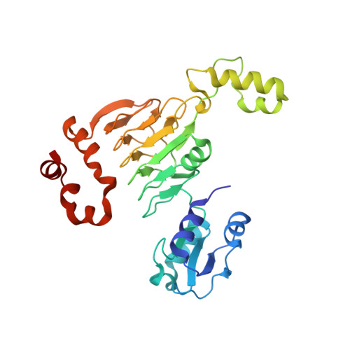
Crystal structure of aminoglycoside resistance enzyme ApmA, complex with neomycin


Experimental Data Snapshot

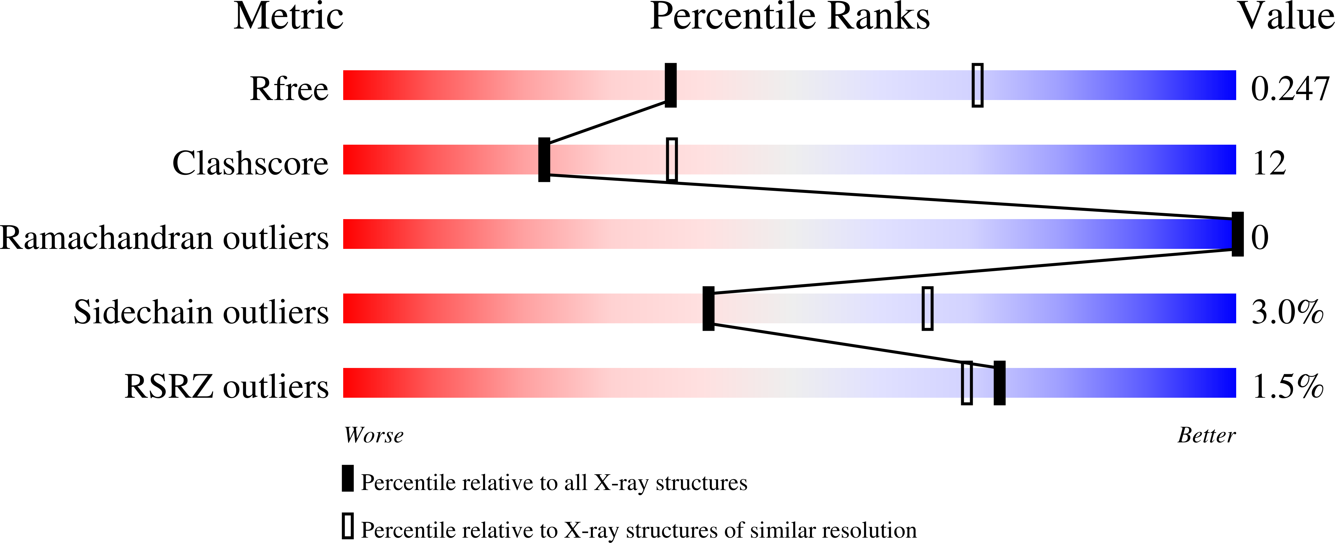
  • Method: X-RAY DIFFRACTION
  • Resolution: 2.83 Å
  • R-Value Free: 0.249 
  • R-Value Work: 0.211 
  • R-Value Observed: 0.213 

Starting Model: experimental
View more details

wwPDB Validation   3D Report Full Report


Ligand Structure Quality Assessment 


This is version 1.4 of the entry. See complete history


Literature

Mechanistic plasticity in ApmA enables aminoglycoside promiscuity for resistance.

Bordeleau, E.Stogios, P.J.Evdokimova, E.Koteva, K.Savchenko, A.Wright, G.D.

(2024) Nat Chem Biol 20: 234-242

  • DOI: https://doi.org/10.1038/s41589-023-01483-3
  • Primary Citation of Related Structures:  
    7UUK, 7UUL, 7UUM, 7UUN, 7UUO

  • PubMed Abstract: 

    The efficacy of aminoglycoside antibiotics is waning due to the acquisition of diverse resistance mechanisms by bacteria. Among the most prevalent are aminoglycoside acetyltransferases (AACs) that inactivate the antibiotics through acetyl coenzyme A-mediated modification. Most AACs are members of the GCN5 superfamily of acyltransferases which lack conserved active site residues that participate in catalysis. ApmA is the first reported AAC belonging to the left-handed β-helix superfamily. These enzymes are characterized by an essential active site histidine that acts as an active site base. Here we show that ApmA confers broad-spectrum aminoglycoside resistance with a molecular mechanism that diverges from other detoxifying left-handed β-helix superfamily enzymes and canonical GCN5 AACs. We find that the active site histidine plays different functions depending on the acetyl-accepting aminoglycoside substrate. This flexibility in the mechanism of a single enzyme underscores the plasticity of antibiotic resistance elements to co-opt protein catalysts in the evolution of drug detoxification.


  • Organizational Affiliation

    David Braley Centre for Antibiotics Discovery, M.G. DeGroote Institute for Infectious Disease Research, Department of Biochemistry and Biomedical Sciences, McMaster University, Hamilton, Ontario, Canada.


Macromolecules
Find similar proteins by:  (by identity cutoff)  |  3D Structure
Entity ID: 1
MoleculeChains Sequence LengthOrganismDetailsImage
Aminocyclitol acetyltransferase ApmA276Staphylococcus aureusMutation(s): 0 
Gene Names: apmA
UniProt
Find proteins for A0A1D0AST6 (Staphylococcus aureus)
Explore A0A1D0AST6 
Go to UniProtKB:  A0A1D0AST6
Entity Groups  
Sequence Clusters30% Identity50% Identity70% Identity90% Identity95% Identity100% Identity
UniProt GroupA0A1D0AST6
Sequence Annotations
Expand
  • Reference Sequence
Experimental Data & Validation

Experimental Data

  • Method: X-RAY DIFFRACTION
  • Resolution: 2.83 Å
  • R-Value Free: 0.249 
  • R-Value Work: 0.211 
  • R-Value Observed: 0.213 
  • Space Group: P 43 3 2
Unit Cell:
Length ( Å )Angle ( ˚ )
a = 133.564α = 90
b = 133.564β = 90
c = 133.564γ = 90
Software Package:
Software NamePurpose
PHENIXrefinement
HKL-3000data reduction
HKL-3000data scaling
PHENIXphasing
PHENIXmodel building
Cootmodel building

Structure Validation

View Full Validation Report


Ligand Structure Quality Assessment 


Entry History & Funding Information

Deposition Data


Funding OrganizationLocationGrant Number
National Institutes of Health/National Institute Of Allergy and Infectious Diseases (NIH/NIAID)United StatesHHSN272201700060C

Revision History  (Full details and data files)

  • Version 1.0: 2022-11-02
    Type: Initial release
  • Version 1.1: 2023-01-25
    Changes: Structure summary
  • Version 1.2: 2023-10-25
    Changes: Data collection, Refinement description
  • Version 1.3: 2023-11-29
    Changes: Database references
  • Version 1.4: 2024-02-14
    Changes: Database references