7WCD

Cryo EM structure of SARS-CoV-2 spike in complex with TAU-2212 mAbs in conformation 4


Experimental Data Snapshot

  • Method: ELECTRON MICROSCOPY
  • Resolution: 3.30 Å
  • Aggregation State: PARTICLE 
  • Reconstruction Method: SINGLE PARTICLE 

wwPDB Validation   3D Report Full Report


This is version 1.1 of the entry. See complete history


Literature

Conformational flexibility in neutralization of SARS-CoV-2 by naturally elicited anti-SARS-CoV-2 antibodies.

Li, R.Mor, M.Ma, B.Clark, A.E.Alter, J.Werbner, M.Lee, J.C.Leibel, S.L.Carlin, A.F.Dessau, M.Gal-Tanamy, M.Croker, B.A.Xiang, Y.Freund, N.T.

(2022) Commun Biol 5: 789-789

  • DOI: https://doi.org/10.1038/s42003-022-03739-5
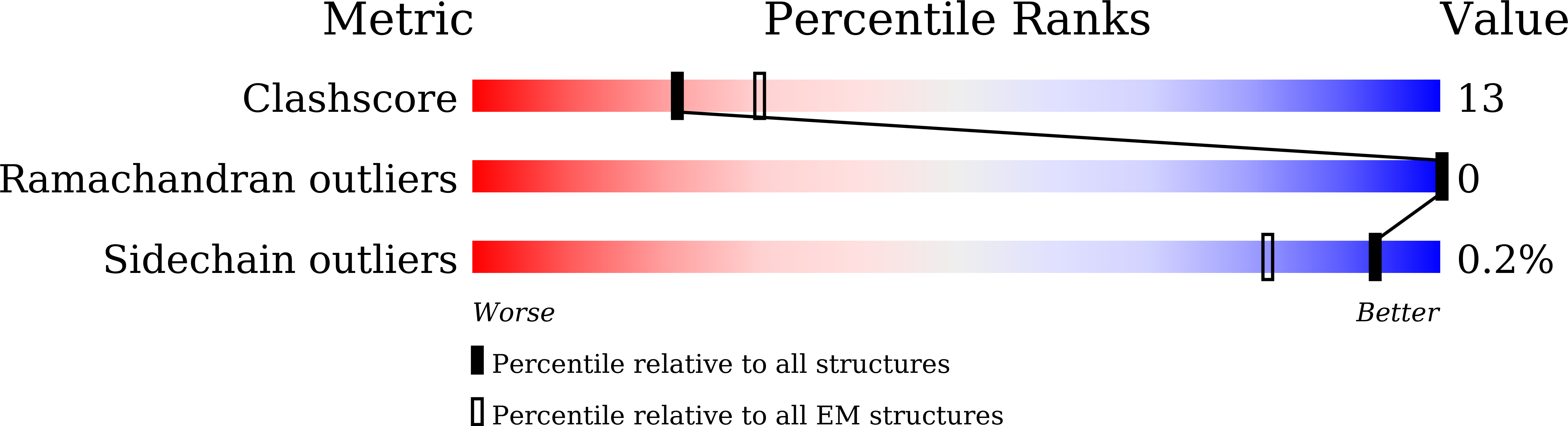
  • Primary Citation of Related Structures:  
    7WBZ, 7WC0, 7WCD

  • PubMed Abstract: 

    As new variants of SARS-CoV-2 continue to emerge, it is important to assess the cross-neutralizing capabilities of antibodies naturally elicited during wild type SARS-CoV-2 infection. In the present study, we evaluate the activity of nine anti-SARS-CoV-2 monoclonal antibodies (mAbs), previously isolated from convalescent donors infected with the Wuhan-Hu-1 strain, against the SARS-CoV-2 variants of concern (VOC) Alpha, Beta, Gamma, Delta and Omicron. By testing an array of mutated spike receptor binding domain (RBD) proteins, cell-expressed spike proteins from VOCs, and neutralization of SARS-CoV-2 VOCs as pseudoviruses, or as the authentic viruses in culture, we show that mAbs directed against the ACE2 binding site (ACE2bs) are more sensitive to viral evolution compared to anti-RBD non-ACE2bs mAbs, two of which retain their potency against all VOCs tested. At the second part of our study, we reveal the neutralization mechanisms at high molecular resolution of two anti-SARS-CoV-2 neutralizing mAbs by structural characterization. We solve the structures of the Delta-neutralizing ACE2bs mAb TAU-2303 with the SARS-CoV-2 spike trimer and RBD at 4.5 Å and 2.42 Å resolutions, respectively, revealing a similar mode of binding to that between the RBD and ACE2. Furthermore, we provide five additional structures (at resolutions of 4.7 Å, 7.3 Å, 6.4 Å, 3.3 Å, and 6.1 Å) of a second antibody, TAU-2212, complexed with the SARS-CoV-2 spike trimer. TAU-2212 binds an exclusively quaternary epitope, and exhibits a unique, flexible mode of neutralization that involves transitioning between five different conformations, with both arms of the antibody recruited for cross linking intra- and inter-spike RBD subunits. Our study provides additional mechanistic understanding about how antibodies neutralize SARS-CoV-2 and its emerging variants and provides insights on the likelihood of reinfections.


  • Organizational Affiliation

    Beijing Advanced Innovation Center for Structural Biology, Beijing Frontier Research Center for Biological Structure, Center for Infectious Disease Research, School of Medicine, Tsinghua University, Beijing, China.


Macromolecules
Find similar proteins by:  (by identity cutoff)  |  3D Structure
Entity ID: 1
MoleculeChains Sequence LengthOrganismDetailsImage
Spike glycoproteinA [auth H],
D [auth J],
G [auth C]
1,237Severe acute respiratory syndrome coronavirus 2Mutation(s): 5 
Gene Names: S2
UniProt
Find proteins for P0DTC2 (Severe acute respiratory syndrome coronavirus 2)
Explore P0DTC2 
Go to UniProtKB:  P0DTC2
Entity Groups  
Sequence Clusters30% Identity50% Identity70% Identity90% Identity95% Identity100% Identity
UniProt GroupP0DTC2
Sequence Annotations
Expand
  • Reference Sequence
Find similar proteins by:  (by identity cutoff)  |  3D Structure
Entity ID: 2
MoleculeChains Sequence LengthOrganismDetailsImage
Heavy chainB [auth E],
E [auth G],
H [auth A]
238Homo sapiensMutation(s): 0 
Entity Groups  
Sequence Clusters30% Identity50% Identity70% Identity90% Identity95% Identity100% Identity
Sequence Annotations
Expand
  • Reference Sequence
Find similar proteins by:  (by identity cutoff)  |  3D Structure
Entity ID: 3
MoleculeChains Sequence LengthOrganismDetailsImage
Light chainC [auth F],
F [auth I],
I [auth B]
214Homo sapiensMutation(s): 0 
Entity Groups  
Sequence Clusters30% Identity50% Identity70% Identity90% Identity95% Identity100% Identity
Sequence Annotations
Expand
  • Reference Sequence
Small Molecules
Ligands 1 Unique
IDChains Name / Formula / InChI Key2D Diagram3D Interactions
NAG
Query on NAG

Download Ideal Coordinates CCD File 
AA [auth J]
BA [auth J]
CA [auth J]
DA [auth J]
EA [auth J]
AA [auth J],
BA [auth J],
CA [auth J],
DA [auth J],
EA [auth J],
FA [auth C],
GA [auth C],
HA [auth C],
IA [auth C],
J [auth H],
JA [auth C],
K [auth H],
KA [auth C],
L [auth H],
LA [auth C],
M [auth H],
MA [auth C],
N [auth H],
NA [auth C],
O [auth H],
OA [auth C],
P [auth H],
PA [auth C],
Q [auth H],
R [auth H],
S [auth H],
T [auth H],
U [auth J],
V [auth J],
W [auth J],
X [auth J],
Y [auth J],
Z [auth J]
2-acetamido-2-deoxy-beta-D-glucopyranose
C8 H15 N O6
OVRNDRQMDRJTHS-FMDGEEDCSA-N
Experimental Data & Validation

Experimental Data

  • Method: ELECTRON MICROSCOPY
  • Resolution: 3.30 Å
  • Aggregation State: PARTICLE 
  • Reconstruction Method: SINGLE PARTICLE 

Structure Validation

View Full Validation Report


Entry History & Funding Information

Deposition Data


Funding OrganizationLocationGrant Number
National Natural Science Foundation of China (NSFC)China--

Revision History  (Full details and data files)

  • Version 1.0: 2022-08-10
    Type: Initial release
  • Version 1.1: 2022-08-17
    Changes: Database references