7Y71

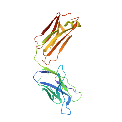
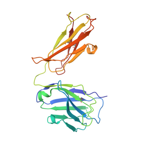
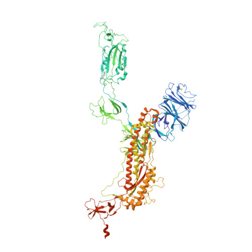
SARS-CoV-2 spike glycoprotein trimer complexed with Fab fragment of anti-RBD antibody E7


Experimental Data Snapshot

  • Method: ELECTRON MICROSCOPY
  • Resolution: 3.12 Å
  • Aggregation State: PARTICLE 
  • Reconstruction Method: SINGLE PARTICLE 

Starting Models: experimental
View more details

wwPDB Validation   3D Report Full Report


This is version 1.2 of the entry. See complete history


Literature

Potent pan huACE2-dependent sarbecovirus neutralizing monoclonal antibodies isolated from a BNT162b2-vaccinated SARS survivor.

Chia, W.N.Tan, C.W.Tan, A.W.K.Young, B.Starr, T.N.Lopez, E.Fibriansah, G.Barr, J.Cheng, S.Yeoh, A.Y.Yap, W.C.Lim, B.L.Ng, T.S.Sia, W.R.Zhu, F.Chen, S.Zhang, J.Kwek, M.S.S.Greaney, A.J.Chen, M.Au, G.G.Paradkar, P.N.Peiris, M.Chung, A.W.Bloom, J.D.Lye, D.Lok, S.Wang, L.F.

(2023) Sci Adv 9: eade3470-eade3470

  • DOI: https://doi.org/10.1126/sciadv.ade3470
  • Primary Citation of Related Structures:  
    7Y71, 7Y72

  • PubMed Abstract: 

    The emergence of severe acute respiratory syndrome coronavirus 2 (SARS-CoV-2) variants of concern such as Omicron hampered efforts in controlling the ongoing coronavirus disease 2019 pandemic due to their ability to escape neutralizing antibodies induced by vaccination or prior infection, highlighting the need to develop broad-spectrum vaccines and therapeutics. Most human monoclonal antibodies (mAbs) reported to date have not demonstrated true pan-sarbecovirus neutralizing breadth especially against animal sarbecoviruses. Here, we report the isolation and characterization of highly potent mAbs targeting the receptor binding domain (RBD) of huACE2-dependent sarbecovirus from a SARS-CoV survivor vaccinated with BNT162b2. Among the six mAbs identified, one (E7) showed better huACE2-dependent sarbecovirus neutralizing potency and breadth than any other mAbs reported to date. Mutagenesis and cryo-electron microscopy studies indicate that these mAbs have a unique RBD contact footprint and that E7 binds to a quaternary structure-dependent epitope.


  • Organizational Affiliation

    Programme in Emerging Infectious Diseases, Duke-NUS Medical School, Singapore, Singapore.


Macromolecules
Find similar proteins by:  (by identity cutoff)  |  3D Structure
Entity ID: 1
MoleculeChains Sequence LengthOrganismDetailsImage
Fab E7 light chainA [auth P]219Homo sapiensMutation(s): 0 
Entity Groups  
Sequence Clusters30% Identity50% Identity70% Identity90% Identity95% Identity100% Identity
Sequence Annotations
Expand
  • Reference Sequence
Find similar proteins by:  (by identity cutoff)  |  3D Structure
Entity ID: 2
MoleculeChains Sequence LengthOrganismDetailsImage
Fab E7 heavy chainB [auth O]221Homo sapiensMutation(s): 0 
Entity Groups  
Sequence Clusters30% Identity50% Identity70% Identity90% Identity95% Identity100% Identity
Sequence Annotations
Expand
  • Reference Sequence
Find similar proteins by:  (by identity cutoff)  |  3D Structure
Entity ID: 3
MoleculeChains Sequence LengthOrganismDetailsImage
Spike glycoproteinC [auth A],
D [auth B],
E [auth C]
1,198Homo sapiensMutation(s): 8 
Gene Names: S2
UniProt
Find proteins for P0DTC2 (Severe acute respiratory syndrome coronavirus 2)
Explore P0DTC2 
Go to UniProtKB:  P0DTC2
Entity Groups  
Sequence Clusters30% Identity50% Identity70% Identity90% Identity95% Identity100% Identity
UniProt GroupP0DTC2
Glycosylation
Glycosylation Sites: 3Go to GlyGen: P0DTC2-1
Sequence Annotations
Expand
  • Reference Sequence
Oligosaccharides

Help

Entity ID: 4
MoleculeChains Length2D Diagram Glycosylation3D Interactions
2-acetamido-2-deoxy-beta-D-glucopyranose-(1-4)-2-acetamido-2-deoxy-beta-D-glucopyranose
F [auth D],
G [auth I],
H [auth J],
I [auth K],
J [auth L],
F [auth D],
G [auth I],
H [auth J],
I [auth K],
J [auth L],
K [auth M],
L [auth N]
2N-Glycosylation
Glycosylation Resources
GlyTouCan:  G42666HT
GlyCosmos:  G42666HT
GlyGen:  G42666HT
Small Molecules
Ligands 1 Unique
IDChains Name / Formula / InChI Key2D Diagram3D Interactions
NAG
Query on NAG

Download Ideal Coordinates CCD File 
M [auth A]
N [auth B]
O [auth B]
P [auth B]
Q [auth B]
M [auth A],
N [auth B],
O [auth B],
P [auth B],
Q [auth B],
R [auth B],
S [auth B],
T [auth B],
U [auth C],
V [auth C],
W [auth C],
X [auth C],
Y [auth C],
Z [auth C]
2-acetamido-2-deoxy-beta-D-glucopyranose
C8 H15 N O6
OVRNDRQMDRJTHS-FMDGEEDCSA-N
Experimental Data & Validation

Experimental Data

  • Method: ELECTRON MICROSCOPY
  • Resolution: 3.12 Å
  • Aggregation State: PARTICLE 
  • Reconstruction Method: SINGLE PARTICLE 
EM Software:
TaskSoftware PackageVersion
RECONSTRUCTIONcryoSPARC3.3.1+220215
MODEL REFINEMENTCoot0.8.9.2-pre
MODEL REFINEMENTPHENIX1.20.1-4487

Structure Validation

View Full Validation Report


Entry History & Funding Information

Deposition Data


Funding OrganizationLocationGrant Number
National Research Foundation (NRF, Singapore)SingaporeNRF2016NRF-NSFC002-013
Other governmentSTPRG-FY19-001
Other governmentCOVID19RF-003
Other governmentCOVID19RF-060
Other governmentMOH-000535/MOH-OFYIRG19nov-0002
Other governmentOFLCG19May-0034
National Health and Medical Research Council (NHMRC, Australia)AustraliaGNT 2008092
Damon Runyon Cancer Research FoundationUnited States--

Revision History  (Full details and data files)

  • Version 1.0: 2023-08-02
    Type: Initial release
  • Version 1.1: 2023-08-09
    Changes: Database references
  • Version 1.2: 2024-10-23
    Changes: Data collection, Refinement description, Structure summary