8ALN | pdb_00008aln

CO-bound [FeFe]-hydrogenase I from Clostridium pasteurianum (CpI) at 1.34 Angstrom


Experimental Data Snapshot

  • Method: X-RAY DIFFRACTION
  • Resolution: 1.34 Å
  • R-Value Free: 
    0.175 (Depositor), 0.180 (DCC) 
  • R-Value Work: 
    0.149 (Depositor), 0.150 (DCC) 
  • R-Value Observed: 
    0.150 (Depositor) 

Starting Model: experimental
View more details

wwPDB Validation   3D Report Full Report


Ligand Structure Quality Assessment 

Created with Raphaël 2.3.0Worse 01 BetterLigand structure goodness of fit to experimental dataBest fitted MHXClick on this verticalbar to view details

This is version 2.1 of the entry. See complete history


Literature

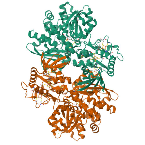
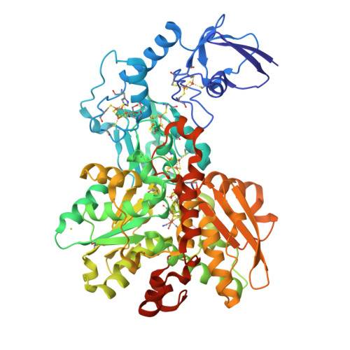
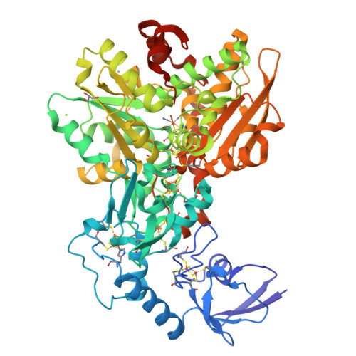
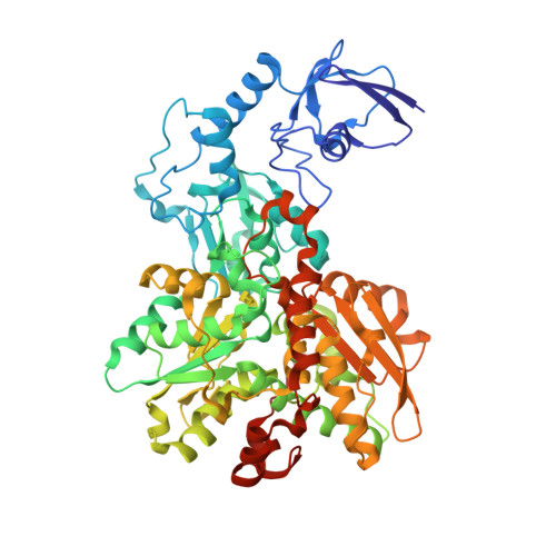
Cyanide Binding to [FeFe]-Hydrogenase Stabilizes the Alternative Configuration of the Proton Transfer Pathway.

Duan, J.Hemschemeier, A.Burr, D.J.Stripp, S.T.Hofmann, E.Happe, T.

(2023) Angew Chem Int Ed Engl 62: e202216903-e202216903

  • DOI: https://doi.org/10.1002/anie.202216903
  • Primary Citation of Related Structures:  
    8AIO, 8AJ6, 8ALN, 8AP2

  • PubMed Abstract: 

    Hydrogenases are H 2 converting enzymes that harbor catalytic cofactors in which iron (Fe) ions are coordinated by biologically unusual carbon monoxide (CO) and cyanide (CN - ) ligands. Extrinsic CO and CN - , however, inhibit hydrogenases. The mechanism by which CN - binds to [FeFe]-hydrogenases is not known. Here, we obtained crystal structures of the CN - -treated [FeFe]-hydrogenase CpI from Clostridium pasteurianum. The high resolution of 1.39 Å allowed us to distinguish intrinsic CN - and CO ligands and to show that extrinsic CN - binds to the open coordination site of the cofactor where CO is known to bind. In contrast to other inhibitors, CN - treated crystals show conformational changes of conserved residues within the proton transfer pathway which could allow a direct proton transfer between E279 and S319. This configuration has been proposed to be vital for efficient proton transfer, but has never been observed structurally.


  • Organizational Affiliation

    Department of Plant Biochemistry, Faculty of Biology and Biotechnology, Photobiotechnology, Ruhr University Bochum, Universitätsstrasse 150, 44801, Bochum, Germany.


Macromolecules
Find similar proteins by:  (by identity cutoff)  |  3D Structure
Entity ID: 1
MoleculeChains Sequence LengthOrganismDetailsImage
Iron hydrogenase 1
A, B
584Clostridium pasteurianumMutation(s): 0 
EC: 1.12.7.2
UniProt
Find proteins for P29166 (Clostridium pasteurianum)
Explore P29166 
Go to UniProtKB:  P29166
Entity Groups  
Sequence Clusters30% Identity50% Identity70% Identity90% Identity95% Identity100% Identity
UniProt GroupP29166
Sequence Annotations
Expand
  • Reference Sequence
Small Molecules
Ligands 6 Unique
IDChains Name / Formula / InChI Key2D Diagram3D Interactions
MHX (Subject of Investigation/LOI)
Query on MHX

Download Ideal Coordinates CCD File 
M [auth A],
X [auth B]
Binuclear [FeFe], di(thiomethyl)amine, carbon monoxide, cyanide cluster (-CO form)
C8 H5 Fe2 N3 O4 S2
HQBPXMLEPWPPEN-UHFFFAOYSA-N
SF4 (Subject of Investigation/LOI)
Query on SF4

Download Ideal Coordinates CCD File 
C [auth A]
D [auth A]
E [auth A]
F [auth A]
N [auth B]
IRON/SULFUR CLUSTER
Fe4 S4
LJBDFODJNLIPKO-UHFFFAOYSA-N
FES
Query on FES

Download Ideal Coordinates CCD File 
G [auth A],
R [auth B]
FE2/S2 (INORGANIC) CLUSTER
Fe2 S2
NIXDOXVAJZFRNF-UHFFFAOYSA-N
GOL
Query on GOL

Download Ideal Coordinates CCD File 
I [auth A],
J [auth A],
T [auth B]
GLYCEROL
C3 H8 O3
PEDCQBHIVMGVHV-UHFFFAOYSA-N
CL
Query on CL

Download Ideal Coordinates CCD File 
W [auth B]CHLORIDE ION
Cl
VEXZGXHMUGYJMC-UHFFFAOYSA-M
MG
Query on MG

Download Ideal Coordinates CCD File 
H [auth A]
K [auth A]
L [auth A]
S [auth B]
U [auth B]
MAGNESIUM ION
Mg
JLVVSXFLKOJNIY-UHFFFAOYSA-N
Experimental Data & Validation

Experimental Data

  • Method: X-RAY DIFFRACTION
  • Resolution: 1.34 Å
  • R-Value Free:  0.175 (Depositor), 0.180 (DCC) 
  • R-Value Work:  0.149 (Depositor), 0.150 (DCC) 
  • R-Value Observed: 0.150 (Depositor) 
Space Group: P 1 21 1
Unit Cell:
Length ( Å )Angle ( ˚ )
a = 89.86α = 90
b = 71.85β = 97.47
c = 103.23γ = 90
Software Package:
Software NamePurpose
PHENIXrefinement
XSCALEdata scaling
PDB_EXTRACTdata extraction
XDSdata reduction
PHENIXphasing

Structure Validation

View Full Validation Report


Ligand Structure Quality Assessment 

Created with Raphaël 2.3.0Worse 01 BetterLigand structure goodness of fit to experimental dataBest fitted MHXClick on this verticalbar to view details

Entry History & Funding Information

Deposition Data


Funding OrganizationLocationGrant Number
German Research Foundation (DFG)Germany--

Revision History  (Full details and data files)

  • Version 1.0: 2022-12-14
    Type: Initial release
  • Version 1.1: 2023-01-25
    Changes: Database references
  • Version 1.2: 2023-02-15
    Changes: Database references
  • Version 2.0: 2023-03-15
    Changes: Atomic model, Author supporting evidence, Data collection, Derived calculations, Non-polymer description, Structure summary
  • Version 2.1: 2024-02-07
    Changes: Data collection, Refinement description