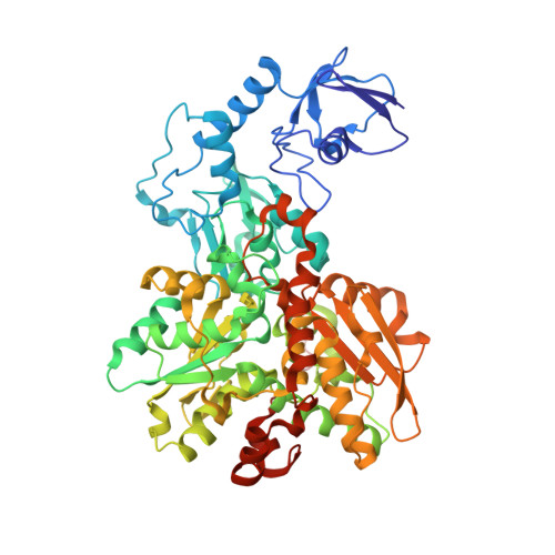
8CJY | pdb_00008cjy

[FeFe]-hydrogenase CpI from Clostridium pasteurianum, variant S357T


Experimental Data Snapshot

  • Method: X-RAY DIFFRACTION
  • Resolution: 1.60 Å
  • R-Value Free: 
    0.187 (Depositor), 0.190 (DCC) 
  • R-Value Work: 
    0.164 (Depositor), 0.160 (DCC) 
  • R-Value Observed: 
    0.165 (Depositor) 

Starting Model: experimental
View more details

wwPDB Validation   3D Report Full Report


Ligand Structure Quality Assessment 

Created with Raphaël 2.3.0Worse 01 BetterLigand structure goodness of fit to experimental dataBest fitted 402Click on this verticalbar to view details

This is version 1.2 of the entry. See complete history


Literature

A Dynamic Water Channel Affects O 2 Stability in [FeFe]-Hydrogenases.

Brocks, C.Das, C.K.Duan, J.Yadav, S.Apfel, U.P.Ghosh, S.Hofmann, E.Winkler, M.Engelbrecht, V.Schafer, L.V.Happe, T.

(2024) ChemSusChem 17: e202301365-e202301365

  • DOI: https://doi.org/10.1002/cssc.202301365
  • Primary Citation of Related Structures:  
    7QHF, 8CJY

  • PubMed Abstract: 

    [FeFe]-hydrogenases are capable of reducing protons at a high rate. However, molecular oxygen (O 2 ) induces the degradation of their catalytic cofactor, the H-cluster, which consists of a cubane [4Fe4S] subcluster (4Fe H ) and a unique diiron moiety (2Fe H ). Previous attempts to prevent O 2 -induced damage have focused on enhancing the protein's sieving effect for O 2 by blocking the hydrophobic gas channels that connect the protein surface and the 2Fe H . In this study, we aimed to block an O 2 diffusion pathway and shield 4Fe H instead. Molecular dynamics (MD) simulations identified a novel water channel (W H ) surrounding the H-cluster. As this hydrophilic path may be accessible for O 2 molecules we applied site-directed mutagenesis targeting amino acids along W H in proximity to 4Fe H to block O 2 diffusion. Protein film electrochemistry experiments demonstrate increased O 2 stabilities for variants G302S and S357T, and MD simulations based on high-resolution crystal structures confirmed an enhanced local sieving effect for O 2 in the environment of the 4Fe H in both cases. The results strongly suggest that, in wild type proteins, O 2 diffuses from the 4Fe H to the 2Fe H . These results reveal new strategies for improving the O 2 stability of [FeFe]-hydrogenases by focusing on the O 2 diffusion network near the active site.


  • Organizational Affiliation

    Faculty of Biology and Biotechnology, Photobiotechnology, Ruhr University Bochum, Universitätsstrasse 150, 44801, Bochum, Germany.


Macromolecules
Find similar proteins by:  (by identity cutoff)  |  3D Structure
Entity ID: 1
MoleculeChains Sequence LengthOrganismDetailsImage
Iron hydrogenase 1
A, B
584Clostridium pasteurianumMutation(s): 1 
EC: 1.12.7.2
UniProt
Find proteins for P29166 (Clostridium pasteurianum)
Explore P29166 
Go to UniProtKB:  P29166
Entity Groups  
Sequence Clusters30% Identity50% Identity70% Identity90% Identity95% Identity100% Identity
UniProt GroupP29166
Sequence Annotations
Expand
  • Reference Sequence
Small Molecules
Ligands 6 Unique
IDChains Name / Formula / InChI Key2D Diagram3D Interactions
402 (Subject of Investigation/LOI)
Query on 402

Download Ideal Coordinates CCD File 
C [auth A],
N [auth B]
dicarbonyl[bis(cyanide-kappaC)]-mu-(iminodimethanethiolatato-1kappaS:2kappaS)-mu-(oxomethylidene)diiron(2+)
C7 H5 Fe2 N3 O3 S2
LJPDYWPSPOWMIB-UHFFFAOYSA-N
SF4 (Subject of Investigation/LOI)
Query on SF4

Download Ideal Coordinates CCD File 
D [auth A]
E [auth A]
F [auth A]
G [auth A]
O [auth B]
IRON/SULFUR CLUSTER
Fe4 S4
LJBDFODJNLIPKO-UHFFFAOYSA-N
FES (Subject of Investigation/LOI)
Query on FES

Download Ideal Coordinates CCD File 
H [auth A],
S [auth B]
FE2/S2 (INORGANIC) CLUSTER
Fe2 S2
NIXDOXVAJZFRNF-UHFFFAOYSA-N
GOL
Query on GOL

Download Ideal Coordinates CCD File 
J [auth A],
U [auth B]
GLYCEROL
C3 H8 O3
PEDCQBHIVMGVHV-UHFFFAOYSA-N
CL
Query on CL

Download Ideal Coordinates CCD File 
L [auth A],
M [auth A],
W [auth B],
X [auth B]
CHLORIDE ION
Cl
VEXZGXHMUGYJMC-UHFFFAOYSA-M
MG
Query on MG

Download Ideal Coordinates CCD File 
I [auth A],
K [auth A],
T [auth B],
V [auth B]
MAGNESIUM ION
Mg
JLVVSXFLKOJNIY-UHFFFAOYSA-N
Experimental Data & Validation

Experimental Data

  • Method: X-RAY DIFFRACTION
  • Resolution: 1.60 Å
  • R-Value Free:  0.187 (Depositor), 0.190 (DCC) 
  • R-Value Work:  0.164 (Depositor), 0.160 (DCC) 
  • R-Value Observed: 0.165 (Depositor) 
Space Group: P 1 21 1
Unit Cell:
Length ( Å )Angle ( ˚ )
a = 89.73α = 90
b = 72.29β = 96.95
c = 103.04γ = 90
Software Package:
Software NamePurpose
XSCALEdata scaling
PHENIXrefinement
XDSdata reduction
PHENIXphasing

Structure Validation

View Full Validation Report


Ligand Structure Quality Assessment 

Created with Raphaël 2.3.0Worse 01 BetterLigand structure goodness of fit to experimental dataBest fitted 402Click on this verticalbar to view details

Entry History & Funding Information

Deposition Data


Funding OrganizationLocationGrant Number
German Research Foundation (DFG)GermanyGRK 2341

Revision History  (Full details and data files)

  • Version 1.0: 2023-10-25
    Type: Initial release
  • Version 1.1: 2023-11-29
    Changes: Database references
  • Version 1.2: 2024-02-21
    Changes: Database references