8CZA

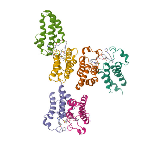
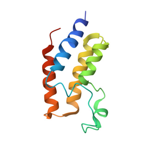
Crystal structure of the first bromodomain (BD1) of human BRDT bound to GXH-IV-075

  • Classification: GENE REGULATION
  • Organism(s): Homo sapiens
  • Expression System: Escherichia coli
  • Mutation(s): No 

  • Deposited: 2022-05-24 Released: 2022-08-03 
  • Deposition Author(s): Chan, A., Schonbrunn, E.
  • Funding Organization(s): National Institutes of Health/Eunice Kennedy Shriver National Institute of Child Health & Human Development (NIH/NICHD)

Experimental Data Snapshot

  • Method: X-RAY DIFFRACTION
  • Resolution: 2.96 Å
  • R-Value Free: 0.268 
  • R-Value Work: 0.204 
  • R-Value Observed: 0.207 

Starting Model: experimental
View more details

wwPDB Validation   3D Report Full Report


Ligand Structure Quality Assessment 


This is version 1.2 of the entry. See complete history

Re-refinement Note

This entry reflects an alternative modeling of the original data in: 7MRE


Literature

Bivalent BET Bromodomain Inhibitors Confer Increased Potency and Selectivity for BRDT via Protein Conformational Plasticity.

Guan, X.Cheryala, N.Karim, R.M.Chan, A.Berndt, N.Qi, J.Georg, G.I.Schonbrunn, E.

(2022) J Med Chem 65: 10441-10458

  • DOI: https://doi.org/10.1021/acs.jmedchem.2c00453
  • Primary Citation of Related Structures:  
    7L9M, 7LA9, 7MR5, 7MR6, 7MR7, 7MR9, 7MRA, 7MRB, 7MRC, 7MRD, 7MRG, 7MRH, 8CZA

  • PubMed Abstract: 

    Bromodomain and extraterminal domain (BET) proteins are important regulators of gene transcription and chromatin remodeling. BET family members BRD4 and BRDT are validated targets for cancer and male contraceptive drug development, respectively. Due to the high structural similarity of the acetyl-lysine binding sites, most reported inhibitors lack intra-BET selectivity. We surmised that protein-protein interactions induced by bivalent inhibitors may differ between BRD4 and BRDT, conferring an altered selectivity profile. Starting from nonselective monovalent inhibitors, we developed cell-active bivalent BET inhibitors with increased activity and selectivity for BRDT. X-ray crystallographic and solution studies revealed unique structural states of BRDT and BRD4 upon interaction with bivalent inhibitors. Varying spacer lengths and symmetric vs unsymmetric connections resulted in the same dimeric states, whereas different chemotypes induced different dimers. The findings indicate that the increased intra-BET selectivity of bivalent inhibitors is due to the differential plasticity of BET bromodomains upon inhibitor-induced dimerization.


  • Organizational Affiliation

    Department of Medicinal Chemistry and Institute for Therapeutics Discovery and Development, College of Pharmacy, University of Minnesota, 717 Delaware Street, Minneapolis, Minnesota 55414, United States.


Macromolecules
Find similar proteins by:  (by identity cutoff)  |  3D Structure
Entity ID: 1
MoleculeChains Sequence LengthOrganismDetailsImage
Bromodomain testis-specific protein
A, B, C, D, E
A, B, C, D, E, F
113Homo sapiensMutation(s): 0 
Gene Names: BRDT
UniProt & NIH Common Fund Data Resources
Find proteins for Q58F21 (Homo sapiens)
Go to UniProtKB:  Q58F21
PHAROS:  Q58F21
GTEx:  ENSG00000137948 
Sequence Annotations
Expand
  • Reference Sequence
Small Molecules
Ligands 2 Unique
IDChains Name / Formula / InChI Key2D Diagram3D Interactions
ZN1 (Subject of Investigation/LOI)
Query on ZN1

Download Ideal Coordinates CCD File 
G [auth B],
H [auth C],
K [auth F]
4-[(4-{4-chloro-3-[(2-methylpropane-2-sulfonyl)amino]anilino}-5-methylpyrimidin-2-yl)amino]-2-fluoro-N-[1-(14-{3-[(2-{3-fluoro-4-[(piperidin-4-yl)carbamoyl]anilino}-5-methylpyrimidin-4-yl)amino]-5-[(2-methylpropane-2-sulfonyl)amino]phenyl}-14-oxo-4,7,10-trioxa-13-azatetradecanan-1-oyl)piperidin-4-yl]benzamide
C64 H82 Cl F2 N15 O11 S2
DPQKUYBPPCXREN-UHFFFAOYSA-N
NA
Query on NA

Download Ideal Coordinates CCD File 
I [auth C],
J [auth D]
SODIUM ION
Na
FKNQFGJONOIPTF-UHFFFAOYSA-N
Binding Affinity Annotations 
IDSourceBinding Affinity
ZN1 BindingDB:  8CZA Kd: min: 0.16, max: 3.1 (nM) from 3 assay(s)
Experimental Data & Validation

Experimental Data

  • Method: X-RAY DIFFRACTION
  • Resolution: 2.96 Å
  • R-Value Free: 0.268 
  • R-Value Work: 0.204 
  • R-Value Observed: 0.207 
  • Space Group: C 2 2 21
Unit Cell:
Length ( Å )Angle ( ˚ )
a = 94.801α = 90
b = 175.737β = 90
c = 143.071γ = 90
Software Package:
Software NamePurpose
PHENIXrefinement
PDB_EXTRACTdata extraction
XDSdata reduction
XSCALEdata scaling
PHENIXphasing

Structure Validation

View Full Validation Report


Ligand Structure Quality Assessment 


Entry History & Funding Information

Deposition Data


Funding OrganizationLocationGrant Number
National Institutes of Health/Eunice Kennedy Shriver National Institute of Child Health & Human Development (NIH/NICHD)United States1U54HD093540

Revision History  (Full details and data files)

  • Version 1.0: 2022-08-03
    Type: Initial release
  • Version 1.1: 2022-08-17
    Changes: Database references
  • Version 1.2: 2023-10-18
    Changes: Data collection, Refinement description